 Movie
Movie Controller


+ Open data

- Basic information
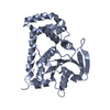
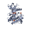
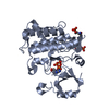
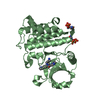
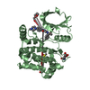
| Entry | Database: PDB / ID: 4e0q | ||||||
|---|---|---|---|---|---|---|---|
| Title | Crystal structure of MPN domain from COP9 signalosome | ||||||
 Components Components | COP9 signalosome complex subunit 6 | ||||||
 Keywords Keywords | UNKNOWN FUNCTION / MPN (Mpr1p and PAD1p N-terminal) domain | ||||||
| Function / homology |  Function and homology information Function and homology informationmale germ-line cyst encapsulation / Formation of TC-NER Pre-Incision Complex / female germ-line stem cell population maintenance / Cargo recognition for clathrin-mediated endocytosis / Neddylation / : / DNA Damage Recognition in GG-NER / multicellular organism development / protein deneddylation / COP9 signalosome ...male germ-line cyst encapsulation / Formation of TC-NER Pre-Incision Complex / female germ-line stem cell population maintenance / Cargo recognition for clathrin-mediated endocytosis / Neddylation / : / DNA Damage Recognition in GG-NER / multicellular organism development / protein deneddylation / COP9 signalosome / oogenesis / germ cell development / protein stabilization / identical protein binding / cytoplasmSimilarity search - Function | ||||||
| Biological species |  Drosophila melanogaster (fruit fly) Drosophila melanogaster (fruit fly) | ||||||
| Method | X-RAY DIFFRACTION / SYNCHROTRON / SAD / Resolution: 2.5 Å | ||||||
 Authors Authors | Zhang, H. / Gao, Z.Q. / Dong, Y.H. | ||||||
 Citation Citation | Journal: Febs Lett. / Year: 2012 Title: The crystal structure of the MPN domain from the COP9 signalosome subunit CSN6 Authors: Zhang, H. / Gao, Z.Q. / Wang, W.J. / Liu, G.F. / Shtykova, E.V. / Xu, J.H. / Li, L.F. / Su, X.D. / Dong, Y.H. | ||||||
| History |
|
- Structure visualization
| Structure viewer | Molecule: MolmilJmol/JSmol |
|---|
- Downloads & links
-Download
| PDBx/mmCIF format | 4e0q.cif.gz | 105.3 KB | Display | PDBx/mmCIF format |
|---|---|---|---|---|
| PDB format | pdb4e0q.ent.gz | 86.9 KB | Display | PDB format |
| PDBx/mmJSON format | 4e0q.json.gz | Tree view | PDBx/mmJSON format | |
| Others |  Other downloads Other downloads |
-Validation report
| Arichive directory | https://data.pdbj.org/pub/pdb/validation_reports/e0/4e0qftp://data.pdbj.org/pub/pdb/validation_reports/e0/4e0q | HTTPS FTP |
|---|
-Related structure data
| Similar structure data |
|---|


-Links
 PDBj
PDBj

- Assembly
| Deposited unit | 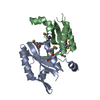
| |||||||||||||||||||||||||||||||||||||||||||||||||
|---|---|---|---|---|---|---|---|---|---|---|---|---|---|---|---|---|---|---|---|---|---|---|---|---|---|---|---|---|---|---|---|---|---|---|---|---|---|---|---|---|---|---|---|---|---|---|---|---|---|---|
| 1 |
| |||||||||||||||||||||||||||||||||||||||||||||||||
| Unit cell |
| |||||||||||||||||||||||||||||||||||||||||||||||||
| Components on special symmetry positions |
| |||||||||||||||||||||||||||||||||||||||||||||||||
| Noncrystallographic symmetry (NCS) | NCS domain:
NCS domain segments:
|
