 Movie
Movie Controller


+ Open data

- Basic information
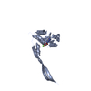
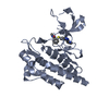
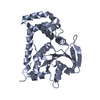
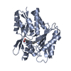
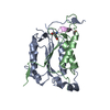
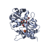
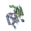
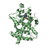
| Entry | Database: PDB / ID: 3bc1 | ||||||
|---|---|---|---|---|---|---|---|
| Title | Crystal Structure of the complex Rab27a-Slp2a | ||||||
 Components Components |
| ||||||
 Keywords Keywords |  SIGNALING PROTEIN/TRANSPORT PROTEIN / Rab27 / GTPase / Rab / signaling protein / GDPNP / Slp2a / Exophilin-4 / GTP-binding / Lipoprotein / Membrane / Methylation / Nucleotide-binding / Prenylation / SIGNALING PROTEIN-TRANSPORT PROTEIN COMPLEX SIGNALING PROTEIN/TRANSPORT PROTEIN / Rab27 / GTPase / Rab / signaling protein / GDPNP / Slp2a / Exophilin-4 / GTP-binding / Lipoprotein / Membrane / Methylation / Nucleotide-binding / Prenylation / SIGNALING PROTEIN-TRANSPORT PROTEIN COMPLEX | ||||||
| Function / homology |  Function and homology information Function and homology informationmultivesicular body organization / cytotoxic T cell degranulation / pigment granule localization / positive regulation of constitutive secretory pathway / pigment granule transport / positive regulation of regulated secretory pathway / melanosome localization / RAB geranylgeranylation / RAB GEFs exchange GTP for GDP on RABs / natural killer cell degranulation ...multivesicular body organization / cytotoxic T cell degranulation / pigment granule localization / positive regulation of constitutive secretory pathway / pigment granule transport / positive regulation of regulated secretory pathway / melanosome localization / RAB geranylgeranylation / RAB GEFs exchange GTP for GDP on RABs / natural killer cell degranulation / exosomal secretion / Weibel-Palade body / melanosome transport / neurexin family protein binding / exocytic vesicle / melanocyte differentiation / multivesicular body sorting pathway / myosin V binding / vesicle docking involved in exocytosis / multivesicular body membrane / complement-dependent cytotoxicity / pigmentation / positive regulation of reactive oxygen species biosynthetic process / phosphatidylserine binding / exocytosis / positive regulation of exocytosis / antigen processing and presentation / photoreceptor outer segment / protein targeting / phosphatase binding / positive regulation of phagocytosis / vesicle-mediated transport / phosphatidylinositol-4,5-bisphosphate binding / Neutrophil degranulation / small monomeric GTPase / secretory granule membrane / secretory granule / intracellular protein transport / small GTPase binding / GDP binding / blood coagulation / melanosome / late endosome / lysosome / apical plasma membrane / protein domain specific binding / GTPase activity / dendrite / positive regulation of gene expression / GTP binding / Golgi apparatus / membrane / plasma membrane / cytoplasmSimilarity search - Function | ||||||
| Biological species |  Mus musculus (house mouse) Mus musculus (house mouse) Homo sapiens (human) Homo sapiens (human) | ||||||
| Method | X-RAY DIFFRACTION / SYNCHROTRON / MOLECULAR REPLACEMENT / Resolution: 1.8 Å | ||||||
 Authors Authors | Chavas, L.M.G. / Ihara, K. / Kawasaki, M. / Wakatsuki, S. | ||||||
 Citation Citation | Journal: Structure / Year: 2008 Title: Elucidation of Rab27 recruitment by its effectors: structure of Rab27a bound to Exophilin4/Slp2-a Authors: Chavas, L.M.G. / Ihara, K. / Kawasaki, M. / Torii, S. / Uejima, T. / Kato, R. / Izumi, T. / Wakatsuki, S. | ||||||
| History |
| ||||||
| Remark 999 | SEQUENCE The sequence database for chain B, F is Q9HCH5-3, isoform 3. |
- Structure visualization
| Structure viewer | Molecule: MolmilJmol/JSmol |
|---|
- Downloads & links
-Download
| PDBx/mmCIF format | 3bc1.cif.gz | 117.9 KB | Display | PDBx/mmCIF format |
|---|---|---|---|---|
| PDB format | pdb3bc1.ent.gz | 89.4 KB | Display | PDB format |
| PDBx/mmJSON format | 3bc1.json.gz | Tree view | PDBx/mmJSON format | |
| Others |  Other downloads Other downloads |
-Validation report
| Arichive directory | https://data.pdbj.org/pub/pdb/validation_reports/bc/3bc1ftp://data.pdbj.org/pub/pdb/validation_reports/bc/3bc1 | HTTPS FTP |
|---|
-Related structure data
| Related structure data |  2if0S S: Starting model for refinement |
|---|---|
| Similar structure data |


-Links
 PDBj
PDBj


- Assembly
| Deposited unit | 
| ||||||||
|---|---|---|---|---|---|---|---|---|---|
| 1 | 
| ||||||||
| 2 | 
| ||||||||
| Unit cell |
|
-Components
| #1: Protein | Mass: 22214.016 Da / Num. of mol.: 2 / Fragment: residues in database 1-193 / Mutation: C123S C188S Source method: isolated from a genetically manipulated source Source: (gene. exp.) Mus musculus (house mouse) / Plasmid: pGEX4T2 / Production host:  Escherichia coli (E. coli) / Strain (production host): BL21(DE3)LysS / References: UniProt: Q9ERI2, small monomeric GTPase Escherichia coli (E. coli) / Strain (production host): BL21(DE3)LysS / References: UniProt: Q9ERI2, small monomeric GTPase#2: Protein | Mass: 7009.934 Da / Num. of mol.: 2 / Fragment: Slp2a Rab-binding domain Source method: isolated from a genetically manipulated source Source: (gene. exp.) Homo sapiens (human) / Plasmid: pGEX4T1 / Production host: Escherichia coli (E. coli) / Strain (production host): BL21(DE3)LysS / References: UniProt: Q9HCH5#3: Chemical | 5'-Guanylyl imidodiphosphate  Mass: 522.196 Da / Num. of mol.: 2 / Source method: obtained synthetically / Formula: C10H17N6O13P3 Mass: 522.196 Da / Num. of mol.: 2 / Source method: obtained synthetically / Formula: C10H17N6O13P3Comment: GppNHp, GMPPNP, energy-carrying molecule analogue*YM #4: Chemical |   Mass: 24.305 Da / Num. of mol.: 2 / Source method: obtained synthetically / Formula: Mg Mass: 24.305 Da / Num. of mol.: 2 / Source method: obtained synthetically / Formula: Mg#5: Water | ChemComp-HOH / | Water Mass: 18.015 Da / Num. of mol.: 351 / Source method: isolated from a natural source / Formula: H2O Mass: 18.015 Da / Num. of mol.: 351 / Source method: isolated from a natural source / Formula: H2O |
|---|
-Experimental details
-Experiment
| Experiment | Method: X-RAY DIFFRACTION / Number of used crystals: 1 |
|---|
- Sample preparation
| Crystal | Density Matthews: 2.05 Å3/Da / Density % sol: 39.89 % |
|---|---|
| Crystal grow | Temperature: 289 K / Method: vapor diffusion, hanging drop / pH: 6.5 Details: Na Citrate, Cacodylate, pH 6.5, VAPOR DIFFUSION, HANGING DROP, temperature 289K |
-Data collection
| Diffraction | Mean temperature: 100 K |
|---|---|
| Diffraction source | Source: SYNCHROTRON / Site: Photon Factory  / Beamline: BL-5A / Wavelength: 1 Å / Beamline: BL-5A / Wavelength: 1 Å |
| Detector | Type: ADSC QUANTUM 315 / Detector: CCD / Date: Oct 10, 2007 / Details: mirrors |
| Radiation | Monochromator: Si(111) / Protocol: SINGLE WAVELENGTH / Monochromatic (M) / Laue (L): M / Scattering type: x-ray |
| Radiation wavelength | Wavelength: 1 Å / Relative weight: 1 |
| Reflection | Resolution: 1.8→30.345 Å / Num. obs: 45347 / % possible obs: 95.7 % / Redundancy: 2.6 % / Biso Wilson estimate: 27.5 Å2 / Rmerge(I) obs: 0.093 / Net I/σ(I): 6.5 |
| Reflection shell | Resolution: 1.8→1.86 Å / Redundancy: 2.5 % / Rmerge(I) obs: 0.634 / Mean I/σ(I) obs: 1.25 / Num. unique all: 4408 / % possible all: 98.6 |
- Processing
| Software |
| ||||||||||||||||||||||||||||||||||||||||||||||||||||||||||||||||||||||||||||||||||||||||||||||||||||||||||||||||||||||||||||||||||||||||||||||||||||||||||||||||||||||||||
|---|---|---|---|---|---|---|---|---|---|---|---|---|---|---|---|---|---|---|---|---|---|---|---|---|---|---|---|---|---|---|---|---|---|---|---|---|---|---|---|---|---|---|---|---|---|---|---|---|---|---|---|---|---|---|---|---|---|---|---|---|---|---|---|---|---|---|---|---|---|---|---|---|---|---|---|---|---|---|---|---|---|---|---|---|---|---|---|---|---|---|---|---|---|---|---|---|---|---|---|---|---|---|---|---|---|---|---|---|---|---|---|---|---|---|---|---|---|---|---|---|---|---|---|---|---|---|---|---|---|---|---|---|---|---|---|---|---|---|---|---|---|---|---|---|---|---|---|---|---|---|---|---|---|---|---|---|---|---|---|---|---|---|---|---|---|---|---|---|---|---|---|
| Refinement | Method to determine structure: MOLECULAR REPLACEMENT Starting model: PDB ENTRY 2IF0 Resolution: 1.8→30 Å / Cor.coef. Fo:Fc: 0.946 / Cor.coef. Fo:Fc free: 0.919 / SU B: 3.422 / SU ML: 0.102 / TLS residual ADP flag: LIKELY RESIDUAL / Cross valid method: THROUGHOUT / ESU R: 0.153 / ESU R Free: 0.145 / Stereochemistry target values: MAXIMUM LIKELIHOOD / Details: HYDROGENS HAVE BEEN ADDED IN THE RIDING POSITIONS
| ||||||||||||||||||||||||||||||||||||||||||||||||||||||||||||||||||||||||||||||||||||||||||||||||||||||||||||||||||||||||||||||||||||||||||||||||||||||||||||||||||||||||||
| Solvent computation | Ion probe radii: 0.8 Å / Shrinkage radii: 0.8 Å / VDW probe radii: 1.2 Å / Solvent model: MASK | ||||||||||||||||||||||||||||||||||||||||||||||||||||||||||||||||||||||||||||||||||||||||||||||||||||||||||||||||||||||||||||||||||||||||||||||||||||||||||||||||||||||||||
| Displacement parameters | Biso mean: 17.074 Å2
| ||||||||||||||||||||||||||||||||||||||||||||||||||||||||||||||||||||||||||||||||||||||||||||||||||||||||||||||||||||||||||||||||||||||||||||||||||||||||||||||||||||||||||
| Refinement step | Cycle: LAST / Resolution: 1.8→30 Å
| ||||||||||||||||||||||||||||||||||||||||||||||||||||||||||||||||||||||||||||||||||||||||||||||||||||||||||||||||||||||||||||||||||||||||||||||||||||||||||||||||||||||||||
| Refine LS restraints |
| ||||||||||||||||||||||||||||||||||||||||||||||||||||||||||||||||||||||||||||||||||||||||||||||||||||||||||||||||||||||||||||||||||||||||||||||||||||||||||||||||||||||||||
| LS refinement shell | Resolution: 1.8→1.847 Å / Total num. of bins used: 20
| ||||||||||||||||||||||||||||||||||||||||||||||||||||||||||||||||||||||||||||||||||||||||||||||||||||||||||||||||||||||||||||||||||||||||||||||||||||||||||||||||||||||||||
| Refinement TLS params. | Method: refined / Refine-ID: X-RAY DIFFRACTION
| ||||||||||||||||||||||||||||||||||||||||||||||||||||||||||||||||||||||||||||||||||||||||||||||||||||||||||||||||||||||||||||||||||||||||||||||||||||||||||||||||||||||||||
| Refinement TLS group |
|
