 Movie
Movie Controller


+ Open data

- Basic information
| Entry | Database: PDB / ID: 4dug | |||||||||
|---|---|---|---|---|---|---|---|---|---|---|
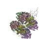
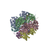
| Title | Crystal Structure of Circadian Clock Protein KaiC E318A Mutant | |||||||||
 Components Components | (Circadian clock protein kinase ... ) x 2 ) x 2 | |||||||||
 Keywords Keywords | CIRCADIAN CLOCK PROTEIN / TRANSFERASE / KaiA / KaiB / ATP-binding / biological rhythms / DNA-binding / metal-binding / nucleotide-binding / phosphoprotein / repressor / Serine/threonine-protein kinase / transcription regulation / auto-kinase / phosphorylation | |||||||||
| Function / homology |  Function and homology information Function and homology informationregulation of phosphorelay signal transduction system / negative regulation of circadian rhythm / entrainment of circadian clock / protein serine/threonine/tyrosine kinase activity / Hydrolases; Acting on acid anhydrides; Acting on acid anhydrides to facilitate cellular and subcellular movement / circadian rhythm / non-specific serine/threonine protein kinase / phosphorylation / protein serine kinase activity / protein serine/threonine kinase activity ...regulation of phosphorelay signal transduction system / negative regulation of circadian rhythm / entrainment of circadian clock / protein serine/threonine/tyrosine kinase activity / Hydrolases; Acting on acid anhydrides; Acting on acid anhydrides to facilitate cellular and subcellular movement / circadian rhythm / non-specific serine/threonine protein kinase / phosphorylation / protein serine kinase activity / protein serine/threonine kinase activity / regulation of DNA-templated transcription / magnesium ion binding / ATP hydrolysis activity / DNA binding / ATP binding / identical protein bindingSimilarity search - Function | |||||||||
| Biological species |  Synechococcus elongatus (bacteria) Synechococcus elongatus (bacteria) | |||||||||
| Method | X-RAY DIFFRACTION / SYNCHROTRON / MOLECULAR REPLACEMENT / Resolution: 3.292 Å | |||||||||
 Authors Authors | Egli, M. / Mori, T. / Pattanayek, R. / Xu, Y. / Qin, X. / Johnson, C.H. | |||||||||
 Citation Citation | Journal: Biochemistry / Year: 2012 Title: Dephosphorylation of the Core Clock Protein KaiC in the Cyanobacterial KaiABC Circadian Oscillator Proceeds via an ATP Synthase Mechanism. Authors: Egli, M. / Mori, T. / Pattanayek, R. / Xu, Y. / Qin, X. / Johnson, C.H. | |||||||||
| History |
|
- Structure visualization
| Structure viewer | Molecule: MolmilJmol/JSmol |
|---|
- Downloads & links
-Download
| PDBx/mmCIF format | 4dug.cif.gz | 601.3 KB | Display | PDBx/mmCIF format |
|---|---|---|---|---|
| PDB format | pdb4dug.ent.gz | 492.3 KB | Display | PDB format |
| PDBx/mmJSON format | 4dug.json.gz | Tree view | PDBx/mmJSON format | |
| Others |  Other downloads Other downloads |
-Validation report
| Arichive directory | https://data.pdbj.org/pub/pdb/validation_reports/du/4dugftp://data.pdbj.org/pub/pdb/validation_reports/du/4dug | HTTPS FTP |
|---|
-Related structure data
| Related structure data |  3dvlS S: Starting model for refinement |
|---|---|
| Similar structure data |


-Links
 PDBj
PDBj
- Assembly
| Deposited unit | 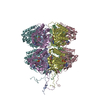
| ||||||||
|---|---|---|---|---|---|---|---|---|---|
| 1 |
| ||||||||
| Unit cell |
|
-Components
-Circadian clock protein kinase ... , 2 types, 6 molecules ABCEFD
| #1: Protein | Mass: 58094.719 Da / Num. of mol.: 5 / Mutation: E318A Source method: isolated from a genetically manipulated source Source: (gene. exp.) Synechococcus elongatus (bacteria) / Strain: PCC 7942 / Gene: kaiC, see0011, Synpcc7942_1216 / Plasmid: PET3 / Production host: Escherichia coli (E. coli) / Strain (production host): Bl21(DE3)References: UniProt: Q79PF4, non-specific serine/threonine protein kinase#2: Protein | | Mass: 58014.734 Da / Num. of mol.: 1 / Mutation: E318A Source method: isolated from a genetically manipulated source Source: (gene. exp.) Synechococcus elongatus (bacteria) / Strain: PCC 7942 / Gene: kaiC, see0011, Synpcc7942_1216 / Plasmid: PET3 / Production host: Escherichia coli (E. coli) / Strain (production host): Bl21(DE3)References: UniProt: Q79PF4, non-specific serine/threonine protein kinase |
|---|
-Non-polymers , 4 types, 94 molecules 


| #3: Chemical | ChemComp-MG /  Mass: 24.305 Da / Num. of mol.: 12 / Source method: obtained synthetically / Formula: Mg Mass: 24.305 Da / Num. of mol.: 12 / Source method: obtained synthetically / Formula: Mg#4: Chemical | ChemComp-ATP / Adenosine triphosphate Mass: 507.181 Da / Num. of mol.: 12 / Source method: obtained synthetically / Formula: C10H16N5O13P3 / Comment: ATP, energy-carrying molecule*YM Mass: 507.181 Da / Num. of mol.: 12 / Source method: obtained synthetically / Formula: C10H16N5O13P3 / Comment: ATP, energy-carrying molecule*YM#5: Chemical | ChemComp-PO4 / | Phosphate Mass: 94.971 Da / Num. of mol.: 1 / Source method: obtained synthetically / Formula: PO4 Mass: 94.971 Da / Num. of mol.: 1 / Source method: obtained synthetically / Formula: PO4#6: Water | ChemComp-HOH / | WaterMass: 18.015 Da / Num. of mol.: 69 / Source method: isolated from a natural source / Formula: H2O |
|---|
-Experimental details
-Experiment
| Experiment | Method: X-RAY DIFFRACTION / Number of used crystals: 1 |
|---|
- Sample preparation
| Crystal | Density Matthews: 2.61 Å3/Da / Density % sol: 52.8 % |
|---|---|
| Crystal grow | Temperature: 291 K / Method: vapor diffusion, hanging drop / pH: 4.8 Details: sodium formate, glycerol, pH 4.8, VAPOR DIFFUSION, HANGING DROP, temperature 291K |
-Data collection
| Diffraction | Mean temperature: 113 K |
|---|---|
| Diffraction source | Source: SYNCHROTRON / Site: APS  / Beamline: 21-ID-F / Wavelength: 0.97872 Å / Beamline: 21-ID-F / Wavelength: 0.97872 Å |
| Detector | Type: MARMOSAIC 225 mm CCD / Detector: CCD / Date: Feb 1, 2010 |
| Radiation | Monochromator: Diamond(111) / Protocol: SINGLE WAVELENGTH / Monochromatic (M) / Laue (L): M / Scattering type: x-ray |
| Radiation wavelength | Wavelength: 0.97872 Å / Relative weight: 1 |
| Reflection | Resolution: 3.292→112.656 Å / Num. all: 55899 / Num. obs: 50551 / % possible obs: 91 % / Observed criterion σ(F): 0 / Observed criterion σ(I): 0 / Redundancy: 8.35 % / Rmerge(I) obs: 0.099 / Net I/σ(I): 10.5 |
| Reflection shell | Resolution: 3.292→3.42 Å / Redundancy: 4 % / Rmerge(I) obs: 0.426 / Mean I/σ(I) obs: 1.42 / Num. unique all: 2791 / % possible all: 51.6 |
- Processing
| Software |
| |||||||||||||||||||||||||||||||||||||||||||||||||||||||||||||||||||||||||||||||||||||||||||||||||||||||||||||||||||||||||||||||||||||
|---|---|---|---|---|---|---|---|---|---|---|---|---|---|---|---|---|---|---|---|---|---|---|---|---|---|---|---|---|---|---|---|---|---|---|---|---|---|---|---|---|---|---|---|---|---|---|---|---|---|---|---|---|---|---|---|---|---|---|---|---|---|---|---|---|---|---|---|---|---|---|---|---|---|---|---|---|---|---|---|---|---|---|---|---|---|---|---|---|---|---|---|---|---|---|---|---|---|---|---|---|---|---|---|---|---|---|---|---|---|---|---|---|---|---|---|---|---|---|---|---|---|---|---|---|---|---|---|---|---|---|---|---|---|---|
| Refinement | Method to determine structure: MOLECULAR REPLACEMENT Starting model: PDB ENTRY 3DVL Resolution: 3.292→30.046 Å / SU ML: 0.4 / Isotropic thermal model: ISOTROPIC / σ(F): 2 / Phase error: 32.32 / Stereochemistry target values: ML
| |||||||||||||||||||||||||||||||||||||||||||||||||||||||||||||||||||||||||||||||||||||||||||||||||||||||||||||||||||||||||||||||||||||
| Solvent computation | Shrinkage radii: 0.86 Å / VDW probe radii: 1.1 Å / Solvent model: FLAT BULK SOLVENT MODEL / Bsol: 36.444 Å2 / ksol: 0.279 e/Å3 | |||||||||||||||||||||||||||||||||||||||||||||||||||||||||||||||||||||||||||||||||||||||||||||||||||||||||||||||||||||||||||||||||||||
| Displacement parameters |
| |||||||||||||||||||||||||||||||||||||||||||||||||||||||||||||||||||||||||||||||||||||||||||||||||||||||||||||||||||||||||||||||||||||
| Refinement step | Cycle: LAST / Resolution: 3.292→30.046 Å
| |||||||||||||||||||||||||||||||||||||||||||||||||||||||||||||||||||||||||||||||||||||||||||||||||||||||||||||||||||||||||||||||||||||
| Refine LS restraints |
| |||||||||||||||||||||||||||||||||||||||||||||||||||||||||||||||||||||||||||||||||||||||||||||||||||||||||||||||||||||||||||||||||||||
| LS refinement shell |
|
