Monochromator: CuK / Protocol: SINGLE WAVELENGTH / Monochromatic (M) / Laue (L): M / Scattering type: x-ray
Radiation wavelength
Wavelength: 1.5418 Å / Relative weight: 1
Reflection
Resolution: 1.7→20 Å / Num. obs: 58454 / % possible obs: 95.9 % / Redundancy: 10.8 % / Rmerge(I) obs: 0.05 / Χ2: 1.183 / Net I/σ(I): 19.6
Reflection shell
Resolution (Å)
Redundancy (%)
Rmerge(I) obs
Num. unique all
Χ2
Diffraction-ID
% possible all
1.7-1.73
10.5
0.565
2758
1.086
1
92.8
1.73-1.76
10.9
0.462
2786
1.117
1
92.9
1.76-1.79
10.9
0.374
2800
1.132
1
93.4
1.79-1.83
11
0.327
2823
1.163
1
93.7
1.83-1.87
11
0.274
2864
1.173
1
94.1
1.87-1.91
11
0.231
2813
1.245
1
94.3
1.91-1.96
11
0.186
2869
1.263
1
94.8
1.96-2.02
11
0.151
2855
1.246
1
94.8
2.02-2.07
11
0.121
2866
1.285
1
95.3
2.07-2.14
11
0.102
2898
1.271
1
95.8
2.14-2.22
11
0.095
2916
1.279
1
95.7
2.22-2.31
10.9
0.085
2896
1.431
1
96.5
2.31-2.41
11
0.071
2932
1.248
1
96.5
2.41-2.54
10.9
0.065
2988
1.247
1
97.1
2.54-2.7
10.9
0.056
2969
1.232
1
97.4
2.7-2.9
10.8
0.049
2969
1.166
1
97.9
2.9-3.2
10.7
0.041
3047
1.098
1
98.2
3.2-3.66
10.4
0.037
3051
1.059
1
98.6
3.66-4.6
10.2
0.034
3094
0.973
1
98.9
4.6-20
9.5
0.032
3260
0.913
1
98.9
-
Processing
Software
Name
Version
Classification
NB
SCALEPACK
datascaling
REFMAC
5.6.0117
refinement
PDB_EXTRACT
3.1
dataextraction
CrystalClear
datacollection
DENZO
datareduction
SHARP
phasing
Refinement
Method to determine structure: SAD / Resolution: 1.7→19.78 Å / Cor.coef. Fo:Fc: 0.956 / Cor.coef. Fo:Fc free: 0.944 / Occupancy max: 1 / Occupancy min: 0.5 / SU B: 1.904 / SU ML: 0.065 / Cross valid method: THROUGHOUT / σ(F): 0 / ESU R: 0.108 / ESU R Free: 0.104 / Stereochemistry target values: MAXIMUM LIKELIHOOD Details: HYDROGENS HAVE BEEN USED IF PRESENT IN THE INPUT U VALUES
Rfactor
Num. reflection
% reflection
Selection details
Rfree
0.233
2993
5.1 %
RANDOM
Rwork
0.2068
-
-
-
obs
0.2081
58426
95.94 %
-
Solvent computation
Ion probe radii: 0.8 Å / Shrinkage radii: 0.8 Å / VDW probe radii: 1.2 Å / Solvent model: MASK
In the structure databanks used in Yorodumi, some data are registered as the other names, "COVID-19 virus" and "2019-nCoV". Here are the details of the virus and the list of structure data.
Jan 31, 2019. EMDB accession codes are about to change! (news from PDBe EMDB page)
EMDB accession codes are about to change! (news from PDBe EMDB page)
The allocation of 4 digits for EMDB accession codes will soon come to an end. Whilst these codes will remain in use, new EMDB accession codes will include an additional digit and will expand incrementally as the available range of codes is exhausted. The current 4-digit format prefixed with “EMD-” (i.e. EMD-XXXX) will advance to a 5-digit format (i.e. EMD-XXXXX), and so on. It is currently estimated that the 4-digit codes will be depleted around Spring 2019, at which point the 5-digit format will come into force.
The EM Navigator/Yorodumi systems omit the EMD- prefix.
Related info.:Q: What is EMD? / ID/Accession-code notation in Yorodumi/EM Navigator
Yorodumi is a browser for structure data from EMDB, PDB, SASBDB, etc.
This page is also the successor to EM Navigator detail page, and also detail information page/front-end page for Omokage search.
The word "yorodu" (or yorozu) is an old Japanese word meaning "ten thousand". "mi" (miru) is to see.
Related info.:EMDB / PDB / SASBDB / Comparison of 3 databanks / Yorodumi Search / Aug 31, 2016. New EM Navigator & Yorodumi / Yorodumi Papers / Jmol/JSmol / Function and homology information / Changes in new EM Navigator and Yorodumi
 Movie
Movie Controller

 Yorodumi
 Open data
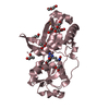
 Basic information
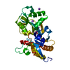
 Components
 Keywords
 PROTEIN BINDING /
  Function and homology information
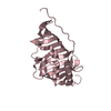
 Authors
 Citation
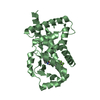
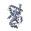
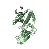
 Structure visualization
 Downloads & links
 Other downloads

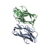


 PDBj
PDBj Assembly


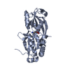
 Mass: 102.046 Da / Num. of mol.: 2 / Source method: obtained synthetically / Formula: C3H2O4
 Mass: 18.015 Da / Num. of mol.: 370 / Source method: isolated from a natural source / Formula: H2O
 Sample preparation
 Processing