 Movie
Movie Controller


+ Open data

- Basic information
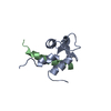
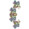
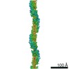
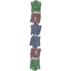
| Entry | Database: PDB / ID: 4d1e | ||||||
|---|---|---|---|---|---|---|---|
| Title | THE CRYSTAL STRUCTURE OF HUMAN MUSCLE ALPHA-ACTININ-2 | ||||||
 Components Components | ALPHA-ACTININ-2 | ||||||
 Keywords Keywords | CONTRACTILE PROTEIN / Z-DISC / CALMODULIN-LIKE DOMAIN / SPECTRIN DOMAIN / ACTIN BINDING DOMAIN / ABD | ||||||
| Function / homology |  Function and homology information Function and homology informationactin filament uncapping / FATZ binding / titin Z domain binding / phospholipase C-activating angiotensin-activated signaling pathway / positive regulation of endocytic recycling / positive regulation of potassium ion transmembrane transporter activity / negative regulation of potassium ion transmembrane transporter activity / positive regulation of cation channel activity / LIM domain binding / negative regulation of protein localization to cell surface ...actin filament uncapping / FATZ binding / titin Z domain binding / phospholipase C-activating angiotensin-activated signaling pathway / positive regulation of endocytic recycling / positive regulation of potassium ion transmembrane transporter activity / negative regulation of potassium ion transmembrane transporter activity / positive regulation of cation channel activity / LIM domain binding / negative regulation of protein localization to cell surface / microspike assembly / postsynaptic actin cytoskeleton / muscle cell development / positive regulation of potassium ion transport / focal adhesion assembly / Assembly and cell surface presentation of NMDA receptors / Striated Muscle Contraction / cardiac muscle cell development / Nephrin family interactions / sarcomere organization / structural constituent of muscle / cortical actin cytoskeleton / Negative regulation of NMDA receptor-mediated neuronal transmission / Unblocking of NMDA receptors, glutamate binding and activation / pseudopodium / postsynaptic density, intracellular component / negative regulation of potassium ion transport / Long-term potentiation / titin binding / phosphatidylinositol-4,5-bisphosphate binding / regulation of membrane potential / Ras activation upon Ca2+ influx through NMDA receptor / cytoskeletal protein binding / nuclear receptor coactivator activity / platelet alpha granule lumen / filopodium / cell projection / actin filament / protein localization to plasma membrane / postsynaptic density membrane / Z disc / actin filament binding / integrin binding / Platelet degranulation / cell junction / actin cytoskeleton organization / RAF/MAP kinase cascade / regulation of apoptotic process / transmembrane transporter binding / dendritic spine / cytoskeleton / cell adhesion / protein domain specific binding / focal adhesion / glutamatergic synapse / calcium ion binding / extracellular exosome / extracellular region / identical protein binding / cytosolSimilarity search - Function | ||||||
| Biological species |  HOMO SAPIENS (human) HOMO SAPIENS (human) | ||||||
| Method | X-RAY DIFFRACTION / SYNCHROTRON / MOLECULAR REPLACEMENT / Resolution: 3.5 Å | ||||||
 Authors Authors | Pinotsis, N. / Salmazo, A. / Sjoeblom, B. / Gkougkoulia, E. / Djinovic-Carugo, K. | ||||||
 Citation Citation | Journal: Cell(Cambridge,Mass.) / Year: 2014 Title: The Structure and Regulation of Human Muscle Alpha-Actinin Authors: Ribeiro Jr, E.A. / Pinotsis, N. / Ghisleni, A. / Salmazo, A. / Konarev, P.V. / Kostan, J. / Sjoeblom, B. / Schreiner, C. / Polyansky, A.A. / Gkougkoulia, E. / Holt, M.R. / Aachmann, F.L. / ...Authors: Ribeiro Jr, E.A. / Pinotsis, N. / Ghisleni, A. / Salmazo, A. / Konarev, P.V. / Kostan, J. / Sjoeblom, B. / Schreiner, C. / Polyansky, A.A. / Gkougkoulia, E. / Holt, M.R. / Aachmann, F.L. / Zagrovic, B. / Bordignon, E. / Pirker, K.F. / Svergun, D.I. / Gautel, M. / Djinovic-Carugo, K. | ||||||
| History |
|
- Structure visualization
| Structure viewer | Molecule: MolmilJmol/JSmol |
|---|
- Downloads & links
-Download
| PDBx/mmCIF format | 4d1e.cif.gz | 365.2 KB | Display | PDBx/mmCIF format |
|---|---|---|---|---|
| PDB format | pdb4d1e.ent.gz | 301.8 KB | Display | PDB format |
| PDBx/mmJSON format | 4d1e.json.gz | Tree view | PDBx/mmJSON format | |
| Others |  Other downloads Other downloads |
-Validation report
| Arichive directory | https://data.pdbj.org/pub/pdb/validation_reports/d1/4d1eftp://data.pdbj.org/pub/pdb/validation_reports/d1/4d1e | HTTPS FTP |
|---|
-Related structure data


-Links
 PDBj
PDBj


- Assembly
| Deposited unit | 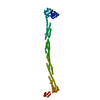
| ||||||||
|---|---|---|---|---|---|---|---|---|---|
| 1 | 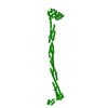
| ||||||||
| Unit cell |
|
-Components
| #1: Protein | / ALPHA-ACTININ SKELETAL MUSCLE ISOFORM 2 / F-ACTIN CROSS-LINKING PROTEIN Mass: 101854.586 Da / Num. of mol.: 1 / Fragment: RESIDUES 19-894 / Mutation: YES Source method: isolated from a genetically manipulated source Details: DELTA 1-18 LYSINE METHYLATION / Source: (gene. exp.) HOMO SAPIENS (human) / Tissue: MUSCLESkeletal muscle / Production host:  ESCHERICHIA COLI (E. coli) / Strain (production host): BL21(DE3) / Variant (production host): ROSETTA PLYSS / References: UniProt: P35609 ESCHERICHIA COLI (E. coli) / Strain (production host): BL21(DE3) / Variant (production host): ROSETTA PLYSS / References: UniProt: P35609 |
|---|
-Experimental details
-Experiment
| Experiment | Method: X-RAY DIFFRACTION / Number of used crystals: 1 |
|---|
- Sample preparation
| Crystal | Density Matthews: 3.48 Å3/Da / Density % sol: 64.65 % / Description: NONE |
|---|---|
| Crystal grow | Details: 0.2 M MG FORMATE 5% PEG SMEAR, 0.01 M EDTA |
-Data collection
| Diffraction | Mean temperature: 100 K |
|---|---|
| Diffraction source | Source: SYNCHROTRON / Site: ESRF  / Beamline: ID23-2 / Wavelength: 0.8726 / Beamline: ID23-2 / Wavelength: 0.8726 |
| Detector | Type: MARRESEARCH / Detector: CCD / Date: Jul 24, 2009 |
| Radiation | Protocol: SINGLE WAVELENGTH / Monochromatic (M) / Laue (L): M / Scattering type: x-ray |
| Radiation wavelength | Wavelength: 0.8726 Å / Relative weight: 1 |
| Reflection | Resolution: 3.5→51.31 Å / Num. obs: 18047 / % possible obs: 99.8 % / Observed criterion σ(I): 1 / Redundancy: 8.5 % / Biso Wilson estimate: 97.7 Å2 / Rmerge(I) obs: 0.2 / Net I/σ(I): 11.2 |
| Reflection shell | Resolution: 3.5→3.83 Å / Redundancy: 8.7 % / Rmerge(I) obs: 1.3 / % possible all: 99.8 |
- Processing
| Software |
| ||||||||||||||||||||||||||||||||||||||||||||||||||||||||||||||||||||||||||||||||||||||||||||||||||||||||||||||||||||||||||||||||||||||||||||||||||||||||||||||||||||||||||||||||||||||||||||||||||||||||
|---|---|---|---|---|---|---|---|---|---|---|---|---|---|---|---|---|---|---|---|---|---|---|---|---|---|---|---|---|---|---|---|---|---|---|---|---|---|---|---|---|---|---|---|---|---|---|---|---|---|---|---|---|---|---|---|---|---|---|---|---|---|---|---|---|---|---|---|---|---|---|---|---|---|---|---|---|---|---|---|---|---|---|---|---|---|---|---|---|---|---|---|---|---|---|---|---|---|---|---|---|---|---|---|---|---|---|---|---|---|---|---|---|---|---|---|---|---|---|---|---|---|---|---|---|---|---|---|---|---|---|---|---|---|---|---|---|---|---|---|---|---|---|---|---|---|---|---|---|---|---|---|---|---|---|---|---|---|---|---|---|---|---|---|---|---|---|---|---|---|---|---|---|---|---|---|---|---|---|---|---|---|---|---|---|---|---|---|---|---|---|---|---|---|---|---|---|---|---|---|---|---|
| Refinement | Method to determine structure: MOLECULAR REPLACEMENT Starting model: PDB ENTRIES 1WKU, 1HCI ,1H8B Resolution: 3.5→51.307 Å / SU ML: 0.36 / σ(F): 1.91 / Phase error: 27.99 / Stereochemistry target values: ML
| ||||||||||||||||||||||||||||||||||||||||||||||||||||||||||||||||||||||||||||||||||||||||||||||||||||||||||||||||||||||||||||||||||||||||||||||||||||||||||||||||||||||||||||||||||||||||||||||||||||||||
| Solvent computation | Shrinkage radii: 1.1 Å / VDW probe radii: 1.3 Å / Solvent model: FLAT BULK SOLVENT MODEL | ||||||||||||||||||||||||||||||||||||||||||||||||||||||||||||||||||||||||||||||||||||||||||||||||||||||||||||||||||||||||||||||||||||||||||||||||||||||||||||||||||||||||||||||||||||||||||||||||||||||||
| Refinement step | Cycle: LAST / Resolution: 3.5→51.307 Å
| ||||||||||||||||||||||||||||||||||||||||||||||||||||||||||||||||||||||||||||||||||||||||||||||||||||||||||||||||||||||||||||||||||||||||||||||||||||||||||||||||||||||||||||||||||||||||||||||||||||||||
| Refine LS restraints |
| ||||||||||||||||||||||||||||||||||||||||||||||||||||||||||||||||||||||||||||||||||||||||||||||||||||||||||||||||||||||||||||||||||||||||||||||||||||||||||||||||||||||||||||||||||||||||||||||||||||||||
| LS refinement shell |
| ||||||||||||||||||||||||||||||||||||||||||||||||||||||||||||||||||||||||||||||||||||||||||||||||||||||||||||||||||||||||||||||||||||||||||||||||||||||||||||||||||||||||||||||||||||||||||||||||||||||||
| Refinement TLS params. | Method: refined / Refine-ID: X-RAY DIFFRACTION
| ||||||||||||||||||||||||||||||||||||||||||||||||||||||||||||||||||||||||||||||||||||||||||||||||||||||||||||||||||||||||||||||||||||||||||||||||||||||||||||||||||||||||||||||||||||||||||||||||||||||||
| Refinement TLS group |
|
