 Movie
Movie Controller


+ Open data

- Basic information
| Entry | Database: PDB / ID: 1hci | ||||||
|---|---|---|---|---|---|---|---|
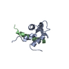
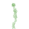
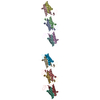
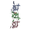
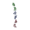
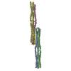
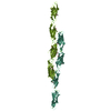
| Title | CRYSTAL STRUCTURE OF THE ROD DOMAIN OF ALPHA-ACTININ | ||||||
 Components Components | ALPHA-ACTININ 2 | ||||||
 Keywords Keywords | TRIPLE-HELIX COILED COIL /  CONTRACTILE PROTEIN / MUSCLE / Z-LINE / ACTIN-BINDING PROTEIN CONTRACTILE PROTEIN / MUSCLE / Z-LINE / ACTIN-BINDING PROTEIN | ||||||
| Function / homology |  Function and homology information Function and homology informationactin filament uncapping / FATZ binding / titin Z domain binding / phospholipase C-activating angiotensin-activated signaling pathway / positive regulation of endocytic recycling / positive regulation of potassium ion transmembrane transporter activity / negative regulation of potassium ion transmembrane transporter activity / positive regulation of cation channel activity / LIM domain binding / negative regulation of protein localization to cell surface ...actin filament uncapping / FATZ binding / titin Z domain binding / phospholipase C-activating angiotensin-activated signaling pathway / positive regulation of endocytic recycling / positive regulation of potassium ion transmembrane transporter activity / negative regulation of potassium ion transmembrane transporter activity / positive regulation of cation channel activity / LIM domain binding / negative regulation of protein localization to cell surface / microspike assembly / postsynaptic actin cytoskeleton / muscle cell development / positive regulation of potassium ion transport / focal adhesion assembly / Assembly and cell surface presentation of NMDA receptors / Striated Muscle Contraction / cardiac muscle cell development / Nephrin family interactions / sarcomere organization / structural constituent of muscle / cortical actin cytoskeleton / Negative regulation of NMDA receptor-mediated neuronal transmission / Unblocking of NMDA receptors, glutamate binding and activation / pseudopodium / postsynaptic density, intracellular component / negative regulation of potassium ion transport / Long-term potentiation / titin binding / phosphatidylinositol-4,5-bisphosphate binding / regulation of membrane potential / Ras activation upon Ca2+ influx through NMDA receptor / cytoskeletal protein binding / nuclear receptor coactivator activity / platelet alpha granule lumen / filopodium / cell projection / actin filament / protein localization to plasma membrane / postsynaptic density membrane / Z disc / actin filament binding / integrin binding / Platelet degranulation / cell junction / actin cytoskeleton organization / RAF/MAP kinase cascade / regulation of apoptotic process / transmembrane transporter binding / dendritic spine / cytoskeleton / cell adhesion / protein domain specific binding / focal adhesion / glutamatergic synapse / calcium ion binding / extracellular exosome / extracellular region / identical protein binding / cytosolSimilarity search - Function | ||||||
| Biological species |  HOMO SAPIENS (human) HOMO SAPIENS (human) | ||||||
| Method | X-RAY DIFFRACTION / SYNCHROTRON / MOLECULAR REPLACEMENT / Resolution: 2.8 Å | ||||||
 Authors Authors | Ylanne, J. / Scheffzek, K. / Young, P. / Saraste, M. | ||||||
 Citation Citation | Journal: Structure / Year: 2001 Title: Crystal Structure of the Alpha-Actinin Rod Reveals an Extensive Torsional Twist Authors: Ylanne, J. / Scheffzek, K. / Young, P. / Saraste, M. | ||||||
| History |
|
- Structure visualization
| Structure viewer | Molecule: MolmilJmol/JSmol |
|---|
- Downloads & links
-Download
| PDBx/mmCIF format | 1hci.cif.gz | 202.1 KB | Display | PDBx/mmCIF format |
|---|---|---|---|---|
| PDB format | pdb1hci.ent.gz | 162.6 KB | Display | PDB format |
| PDBx/mmJSON format | 1hci.json.gz | Tree view | PDBx/mmJSON format | |
| Others |  Other downloads Other downloads |
-Validation report
| Arichive directory | https://data.pdbj.org/pub/pdb/validation_reports/hc/1hciftp://data.pdbj.org/pub/pdb/validation_reports/hc/1hci | HTTPS FTP |
|---|
-Related structure data
| Related structure data |  1quuS S: Starting model for refinement |
|---|---|
| Similar structure data |
-Links
 PDBj
PDBj


- Assembly
| Deposited unit | 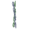
| ||||||||
|---|---|---|---|---|---|---|---|---|---|
| 1 |
| ||||||||
| Unit cell |
|
-Components
| #1: Protein | Mass: 56211.324 Da / Num. of mol.: 2 Fragment: SPECTRIN-LIKE REPEATS 1,2,3, AND 4 - AMINO ACIDS 274 - 746 Source method: isolated from a genetically manipulated source Source: (gene. exp.) HOMO SAPIENS (human) / Tissue: SKELETAL MUSCLE / Plasmid: PET 8C / Cellular location (production host): CYTOPLASM / Production host:  ESCHERICHIA COLI (E. coli) / Strain (production host): BL21(DE3) / References: UniProt: P35609 ESCHERICHIA COLI (E. coli) / Strain (production host): BL21(DE3) / References: UniProt: P35609#2: Water | ChemComp-HOH / | Water Mass: 18.015 Da / Num. of mol.: 16 / Source method: isolated from a natural source / Formula: H2O Mass: 18.015 Da / Num. of mol.: 16 / Source method: isolated from a natural source / Formula: H2OSequence details | PDB CONTAINS N-TERM GLY SER SER FROM THE VECTOR | |
|---|
-Experimental details
-Experiment
| Experiment | Method: X-RAY DIFFRACTION / Number of used crystals: 1 |
|---|
- Sample preparation
| Crystal | Density % sol: 80.3 % | ||||||||||||||||||||||||||||||
|---|---|---|---|---|---|---|---|---|---|---|---|---|---|---|---|---|---|---|---|---|---|---|---|---|---|---|---|---|---|---|---|
| Crystal grow | pH: 8.5 / Details: 0.9 M (NH4)2SO4, 0.1 M TRISHCL PH 8.5 | ||||||||||||||||||||||||||||||
| Crystal grow | *PLUS Temperature: 20 ℃ / pH: 8.5 / Method: vapor diffusion, hanging drop | ||||||||||||||||||||||||||||||
| Components of the solutions | *PLUS
|
-Data collection
| Diffraction | Mean temperature: 100 K |
|---|---|
| Diffraction source | Source: SYNCHROTRON / Site: ESRF  / Beamline: ID14-2 / Wavelength: 0.933 / Beamline: ID14-2 / Wavelength: 0.933 |
| Detector | Type: ADSC CCD / Detector: CCD / Date: Apr 2, 2001 |
| Radiation | Protocol: SINGLE WAVELENGTH / Monochromatic (M) / Laue (L): M / Scattering type: x-ray |
| Radiation wavelength | Wavelength: 0.933 Å / Relative weight: 1 |
| Reflection | Resolution: 2.8→82.78 Å / Num. obs: 63392 / % possible obs: 98.7 % / Observed criterion σ(I): 0 / Redundancy: 2.45 % / Biso Wilson estimate: 21.6 Å2 / Rmerge(I) obs: 0.082 / Net I/σ(I): 7.6 |
| Reflection shell | Resolution: 2.8→2.9 Å / Redundancy: 1.78 % / Rmerge(I) obs: 0.261 / Mean I/σ(I) obs: 2.28 / % possible all: 99 |
| Reflection | *PLUS Num. measured all: 155820 |
| Reflection shell | *PLUS % possible obs: 99 % |
- Processing
| Software |
| ||||||||||||||||||||||||||||||||||||||||||||||||||||||||||||
|---|---|---|---|---|---|---|---|---|---|---|---|---|---|---|---|---|---|---|---|---|---|---|---|---|---|---|---|---|---|---|---|---|---|---|---|---|---|---|---|---|---|---|---|---|---|---|---|---|---|---|---|---|---|---|---|---|---|---|---|---|---|
| Refinement | Method to determine structure: MOLECULAR REPLACEMENT Starting model: PDB ENTRY 1QUU Resolution: 2.8→82.78 Å / Rfactor Rfree error: 0.004 / Data cutoff high absF: 3989928.9 / Cross valid method: THROUGHOUT / σ(F): 0 Stereochemistry target values: MAXIMUM LIKEHOOD USING AMPLITUDES
| ||||||||||||||||||||||||||||||||||||||||||||||||||||||||||||
| Solvent computation | Solvent model: FLAT MODEL / Bsol: 58.9267 Å2 / ksol: 0.367476 e/Å3 | ||||||||||||||||||||||||||||||||||||||||||||||||||||||||||||
| Displacement parameters | Biso mean: 71.8 Å2
| ||||||||||||||||||||||||||||||||||||||||||||||||||||||||||||
| Refine analyze |
| ||||||||||||||||||||||||||||||||||||||||||||||||||||||||||||
| Refinement step | Cycle: LAST / Resolution: 2.8→82.78 Å
| ||||||||||||||||||||||||||||||||||||||||||||||||||||||||||||
| Refine LS restraints |
| ||||||||||||||||||||||||||||||||||||||||||||||||||||||||||||
| LS refinement shell | Resolution: 2.8→2.98 Å / Rfactor Rfree error: 0.014 / Total num. of bins used: 6
| ||||||||||||||||||||||||||||||||||||||||||||||||||||||||||||
| Xplor file |
| ||||||||||||||||||||||||||||||||||||||||||||||||||||||||||||
| Software | *PLUS Name: CNS / Version: 0.9 / Classification: refinement | ||||||||||||||||||||||||||||||||||||||||||||||||||||||||||||
| Refinement | *PLUS Rfactor obs: 0.27 / Rfactor Rwork: 0.27 | ||||||||||||||||||||||||||||||||||||||||||||||||||||||||||||
| Solvent computation | *PLUS | ||||||||||||||||||||||||||||||||||||||||||||||||||||||||||||
| Displacement parameters | *PLUS | ||||||||||||||||||||||||||||||||||||||||||||||||||||||||||||
| Refine LS restraints | *PLUS
|
