- PDB-4ct0: Crystal Structure of Mouse Cryptochrome1 in Complex with Period2 -
+
Open data
ID or keywords:
Loading...
-
Basic information
Entry
Database: PDB / ID: 4ct0
Title
Crystal Structure of Mouse Cryptochrome1 in Complex with Period2
Components
CRYPTOCHROME-1
PERIOD CIRCADIAN PROTEIN HOMOLOG 2
Keywords
CIRCADIAN CLOCK PROTEIN / CRYPTOCHROME-PERIOD COMPLEX / CRYPTOCHROME INTERACTIONS / ZINC INTERFACE / DISULFIDE BOND / REDOX REGULATION
Function / homology
Function and homology information
regulation of glutamate uptake involved in transmission of nerve impulse / circadian regulation of translation / negative regulation of termination of DNA-templated transcription / negative regulation of glucocorticoid secretion / negative regulation of fat cell proliferation / negative regulation of glucocorticoid receptor signaling pathway / negative regulation of transcription regulatory region DNA binding / negative regulation of circadian rhythm / lactate biosynthetic process / negative regulation of G protein-coupled receptor signaling pathway ...regulation of glutamate uptake involved in transmission of nerve impulse / circadian regulation of translation / negative regulation of termination of DNA-templated transcription / negative regulation of glucocorticoid secretion / negative regulation of fat cell proliferation / negative regulation of glucocorticoid receptor signaling pathway / negative regulation of transcription regulatory region DNA binding / negative regulation of circadian rhythm / lactate biosynthetic process / negative regulation of G protein-coupled receptor signaling pathway / histone methyltransferase binding / lipid storage / regulation of DNA damage checkpoint / pre-mRNA binding / glycogen biosynthetic process / RNA polymerase binding / response to glucagon / regulation of gluconeogenesis / entrainment of circadian clock by photoperiod / E-box binding / photoreceptor activity / regulation of insulin secretion / response to light stimulus / regulation of vasoconstriction / white fat cell differentiation / regulation of neurogenesis / phosphatase binding / negative regulation of gluconeogenesis / signal transduction in response to DNA damage / positive regulation of gluconeogenesis / negative regulation of protein ubiquitination / FAD binding / transcription corepressor binding / fatty acid metabolic process / response to activity / positive regulation of protein ubiquitination / response to ischemia / gluconeogenesis / nuclear receptor binding / circadian regulation of gene expression / response to insulin / regulation of circadian rhythm / kinase binding / circadian rhythm / histone deacetylase binding / glucose homeostasis / positive regulation of cold-induced thermogenesis / double-stranded DNA binding / DNA-binding transcription factor binding / transcription coactivator activity / transcription cis-regulatory region binding / regulation of cell cycle / chromatin remodeling / RNA polymerase II cis-regulatory region sequence-specific DNA binding / negative regulation of DNA-templated transcription / protein kinase binding / perinuclear region of cytoplasm / negative regulation of transcription by RNA polymerase II / mitochondrion / DNA binding / nucleoplasm / identical protein binding / nucleus / cytosol / cytoplasm Similarity search - Function
: / Period circadian protein homolog 3-like, PAS-A domain / Period circadian-like, C-terminal / Period protein 2/3C-terminal region / DNA Cyclobutane Dipyrimidine Photolyase, subunit A; domain 3 / DNA Cyclobutane Dipyrimidine Photolyase, subunit A, domain 3 / Serine Threonine Protein Phosphatase 5, Tetratricopeptide repeat - #80 / Cryptochrome/DNA photolyase class 1 / Cryptochrome/DNA photolyase, FAD-binding domain / FAD binding domain of DNA photolyase ...: / Period circadian protein homolog 3-like, PAS-A domain / Period circadian-like, C-terminal / Period protein 2/3C-terminal region / DNA Cyclobutane Dipyrimidine Photolyase, subunit A; domain 3 / DNA Cyclobutane Dipyrimidine Photolyase, subunit A, domain 3 / Serine Threonine Protein Phosphatase 5, Tetratricopeptide repeat - #80 / Cryptochrome/DNA photolyase class 1 / Cryptochrome/DNA photolyase, FAD-binding domain / FAD binding domain of DNA photolyase / PAS fold-3 / PAS fold / DNA photolyase, N-terminal / Cryptochrome/photolyase, N-terminal domain superfamily / DNA photolyase / Photolyase/cryptochrome alpha/beta domain profile. / Cryptochrome/DNA photolyase, FAD-binding domain-like superfamily / HUPs / PAS domain / PAS repeat profile. / Rossmann-like alpha/beta/alpha sandwich fold / PAS domain / PAS domain superfamily / Serine Threonine Protein Phosphatase 5, Tetratricopeptide repeat / Alpha Horseshoe / Rossmann fold / Orthogonal Bundle / 3-Layer(aba) Sandwich / Mainly Alpha / Alpha Beta Similarity search - Domain/homology
Resolution: 2.45→45.496 Å / SU ML: 0.28 / σ(F): 1.34 / Phase error: 24.45 / Stereochemistry target values: ML Details: CHAIN A RESIDUES 1-2 AND 233-240, CHAIN B RESIDUES 1215-1252 ARE DISORDERED
Rfactor
Num. reflection
% reflection
Rfree
0.2331
1695
5 %
Rwork
0.186
-
-
obs
0.1884
33913
99.98 %
Solvent computation
Shrinkage radii: 0.9 Å / VDW probe radii: 1.11 Å / Solvent model: FLAT BULK SOLVENT MODEL
Displacement parameters
Biso mean: 69.8 Å2
Refinement step
Cycle: LAST / Resolution: 2.45→45.496 Å
Protein
Nucleic acid
Ligand
Solvent
Total
Num. atoms
4570
0
21
175
4766
Refine LS restraints
Refine-ID
Type
Dev ideal
Number
X-RAY DIFFRACTION
f_bond_d
0.009
4719
X-RAY DIFFRACTION
f_angle_d
1.125
6421
X-RAY DIFFRACTION
f_dihedral_angle_d
14.681
1692
X-RAY DIFFRACTION
f_chiral_restr
0.079
690
X-RAY DIFFRACTION
f_plane_restr
0.005
826
LS refinement shell
Resolution (Å)
Rfactor Rfree
Num. reflection Rfree
Rfactor Rwork
Num. reflection Rwork
Refine-ID
% reflection obs (%)
2.45-2.5221
0.268
139
0.2292
2642
X-RAY DIFFRACTION
100
2.5221-2.6035
0.2786
138
0.2257
2620
X-RAY DIFFRACTION
100
2.6035-2.6965
0.2845
139
0.2313
2640
X-RAY DIFFRACTION
100
2.6965-2.8045
0.2967
140
0.2312
2632
X-RAY DIFFRACTION
100
2.8045-2.9321
0.2591
139
0.223
2662
X-RAY DIFFRACTION
100
2.9321-3.0867
0.28
140
0.2159
2638
X-RAY DIFFRACTION
100
3.0867-3.28
0.2517
138
0.2154
2663
X-RAY DIFFRACTION
100
3.28-3.5332
0.2807
141
0.2039
2677
X-RAY DIFFRACTION
100
3.5332-3.8885
0.1908
141
0.1728
2670
X-RAY DIFFRACTION
100
3.8885-4.4508
0.198
142
0.1607
2720
X-RAY DIFFRACTION
100
4.4508-5.6059
0.2341
146
0.1745
2751
X-RAY DIFFRACTION
100
5.6059-45.5043
0.223
152
0.1774
2903
X-RAY DIFFRACTION
100
Refinement TLS params.
Method: refined / Refine-ID: X-RAY DIFFRACTION
ID
L11 (°2)
L12 (°2)
L13 (°2)
L22 (°2)
L23 (°2)
L33 (°2)
S11 (Å °)
S12 (Å °)
S13 (Å °)
S21 (Å °)
S22 (Å °)
S23 (Å °)
S31 (Å °)
S32 (Å °)
S33 (Å °)
T11 (Å2)
T12 (Å2)
T13 (Å2)
T22 (Å2)
T23 (Å2)
T33 (Å2)
Origin x (Å)
Origin y (Å)
Origin z (Å)
1
3.7448
-1.9902
2.7913
3.0726
-0.7236
4.7958
-0.0788
0.1666
0.1428
-0.1719
-0.4301
-0.6843
-0.3135
1.1317
0.3386
0.7352
-0.138
0.104
0.4984
0.1759
0.4612
-6.8148
1.6143
-25.3233
2
2.7903
-0.0997
-0.4915
2.3933
-0.6444
4.2595
0.102
0.2062
0.0895
-0.3238
-0.2236
-0.5129
-0.4075
1.235
0.0013
0.3846
-0.0826
0.0392
0.5347
0.1116
0.4231
-2.4993
-4.3168
-15.4485
3
1.9718
-0.7901
0.5576
3.5969
-0.7363
3.4739
-0.34
-0.0071
0.4431
-0.4271
0.031
0.0207
-1.2745
1.1929
0.2006
1.1174
-0.0995
-0.1205
0.049
0.1474
0.4753
-16.0825
10.2614
-19.8389
4
2.2455
-0.0911
0.6176
2.4241
-1.0496
4.7773
0.0501
-0.3703
0.0325
-0.4112
0.3281
0.6894
-0.4203
-1.5821
-0.0435
0.4257
0.1114
-0.1646
0.6928
0.2035
0.5294
-37.0405
-6.7716
-16.2356
5
1.325
-0.5439
-0.8939
6.9259
0.9245
1.4763
0.2837
-0.0891
-0.0285
-0.1484
0.223
0.393
0.5465
0.0422
0.1294
1.1803
-0.044
-0.1379
-0.1534
-0.0604
0.6593
-23.8184
-16.6474
-31.1965
6
7.4789
4.2966
2.6622
5.3468
4.3459
4.1827
-0.4233
0.4792
-0.0445
-0.8002
0.248
-0.0178
-0.4968
0.0008
-0.0604
1.3457
-0.4604
-0.4606
0.5464
0.0627
0.7466
-34.8575
-22.1355
-29.9835
7
3.7193
1.1991
1.764
0.404
0.6514
4.7536
0.1301
0.8106
-0.0992
-0.4515
-0.2145
0.1684
-0.0473
0.0333
-0.2017
0.7884
-0.3267
-1.0537
1.233
0.4275
0.6423
-42.965
-17.6011
-33.0299
8
7.1786
1.4764
4.1165
6.5154
4.223
4.2044
0.134
-0.2928
-0.1553
-0.1741
-0.0514
0.3983
0.2633
-1.0237
-0.0614
1.026
-0.4852
-0.3104
1.4205
0.2482
0.7409
-47.9581
-20.763
-21.3002
9
4.0293
-1.9065
-1.3786
5.5549
-0.0546
4.0863
0.0602
-0.7394
-0.7246
1.1379
0.4768
0.5962
0.3478
-2.0188
-0.6297
0.7334
-0.3736
-0.1824
1.0914
0.3626
0.489
-39.0134
-26.2113
-2.6714
10
6.5858
-3.423
-0.9503
3.5356
-0.1504
1.8236
-0.206
-0.0454
-0.1135
-0.0464
-0.0912
-0.6168
-0.0152
-0.2455
-0.1573
-0.08
-0.0489
0.0471
1.0149
0.1333
0.4803
-26.7003
-16.5952
4.6841
11
9.0866
1.8171
0.0182
9.4605
-1.4385
3.0272
0.0297
-1.1452
0.2281
1.7443
0.55
0.0906
-1.0007
-2.8422
-0.6802
0.8415
0.0139
0.0456
1.157
0.1364
0.5153
-26.3848
-5.3918
10.0847
12
0.8791
1.8001
1.2812
4.4954
2.4209
1.9307
-0.26
-1.8072
-0.1912
2.0501
-0.8751
1.5703
0.571
-0.6472
0.6855
0.8065
0.0615
0.2371
1.6627
0.1038
0.674
-34.7204
-7.3664
7.8235
13
5.4278
0.5517
-4.2581
2.7438
-0.4976
3.3392
-0.0358
0.2959
-0.2001
-0.0772
-0.1634
0.4241
0.0002
-0.6666
-0.1515
0.0148
0.1243
0.3401
1.999
0.5822
0.8205
-43.2201
-8.8425
2.4935
14
6.3701
-7.5888
6.0662
9.1672
-7.2203
5.7798
0.1446
-2.3788
-0.672
-0.0338
1.3069
1.9515
-0.0911
-4.4462
-1.2969
0.6551
0.1731
-0.1958
2.0898
0.171
0.5675
-51.2828
-3.9213
-9.5902
15
0.8036
0.6982
0.2012
0.8681
0.4791
0.4326
0.0458
0.0165
-0.6257
-0.4082
0.2116
-0.3225
0.6186
0.1898
0.5903
1.1916
0.0487
-0.2954
0.1788
0.1688
0.8644
-18.9413
-14.875
-32.7366
Refinement TLS group
ID
Refine-ID
Refine TLS-ID
Selection details
1
X-RAY DIFFRACTION
1
CHAINAAND (RESID3THROUGH69 )
2
X-RAY DIFFRACTION
2
CHAINAAND (RESID70THROUGH149 )
3
X-RAY DIFFRACTION
3
CHAINAAND (RESID150THROUGH283 )
4
X-RAY DIFFRACTION
4
CHAINAAND (RESID284THROUGH495 )
5
X-RAY DIFFRACTION
5
CHAINBAND (RESID1132THROUGH1141 )
6
X-RAY DIFFRACTION
6
CHAINBAND (RESID1142THROUGH1146 )
7
X-RAY DIFFRACTION
7
CHAINBAND (RESID1147THROUGH1151 )
8
X-RAY DIFFRACTION
8
CHAINBAND (RESID1152THROUGH1159 )
9
X-RAY DIFFRACTION
9
CHAINBAND (RESID1160THROUGH1177 )
10
X-RAY DIFFRACTION
10
CHAINBAND (RESID1178THROUGH1182 )
11
X-RAY DIFFRACTION
11
CHAINBAND (RESID1183THROUGH1192 )
12
X-RAY DIFFRACTION
12
CHAINBAND (RESID1193THROUGH1198 )
13
X-RAY DIFFRACTION
13
CHAINBAND (RESID1199THROUGH1208 )
14
X-RAY DIFFRACTION
14
CHAINBAND (RESID1209THROUGH1214 )
15
X-RAY DIFFRACTION
15
CHAIN B AND (RESID -12 THROUGH 0)
+
About Yorodumi
-
News
-
Feb 9, 2022. New format data for meta-information of EMDB entries
New format data for meta-information of EMDB entries
Version 3 of the EMDB header file is now the official format.
The previous official version 1.9 will be removed from the archive.
In the structure databanks used in Yorodumi, some data are registered as the other names, "COVID-19 virus" and "2019-nCoV". Here are the details of the virus and the list of structure data.
Jan 31, 2019. EMDB accession codes are about to change! (news from PDBe EMDB page)
EMDB accession codes are about to change! (news from PDBe EMDB page)
The allocation of 4 digits for EMDB accession codes will soon come to an end. Whilst these codes will remain in use, new EMDB accession codes will include an additional digit and will expand incrementally as the available range of codes is exhausted. The current 4-digit format prefixed with “EMD-” (i.e. EMD-XXXX) will advance to a 5-digit format (i.e. EMD-XXXXX), and so on. It is currently estimated that the 4-digit codes will be depleted around Spring 2019, at which point the 5-digit format will come into force.
The EM Navigator/Yorodumi systems omit the EMD- prefix.
Related info.:Q: What is EMD? / ID/Accession-code notation in Yorodumi/EM Navigator
Yorodumi is a browser for structure data from EMDB, PDB, SASBDB, etc.
This page is also the successor to EM Navigator detail page, and also detail information page/front-end page for Omokage search.
The word "yorodu" (or yorozu) is an old Japanese word meaning "ten thousand". "mi" (miru) is to see.
Related info.:EMDB / PDB / SASBDB / Comparison of 3 databanks / Yorodumi Search / Aug 31, 2016. New EM Navigator & Yorodumi / Yorodumi Papers / Jmol/JSmol / Function and homology information / Changes in new EM Navigator and Yorodumi
 Movie
Movie Controller


 Open data
 Basic information
 Components
 Keywords
 CIRCADIAN CLOCK PROTEIN / CRYPTOCHROME-PERIOD COMPLEX / CRYPTOCHROME INTERACTIONS / ZINC INTERFACE /
  Function and homology information
 Authors
 Citation
 Structure visualization
 Downloads & links
 Other downloads
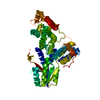

 PDBj
PDBj


 Assembly


 Mass: 65.409 Da / Num. of mol.: 1 / Source method: obtained synthetically / Formula: Zn
 Mass: 35.453 Da / Num. of mol.: 1 / Source method: obtained synthetically / Formula: Cl
 Mass: 282.331 Da / Num. of mol.: 1 / Source method: obtained synthetically / Formula: C12H26O7 / Comment: precipitant*YM
 Sample preparation
 / Beamline: X10SA / Wavelength: 0.9716
  Processing