| Entry | Database: PDB / ID: 3zyz
|
|---|
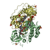
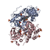
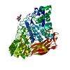
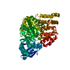
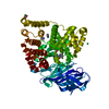
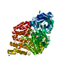
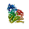
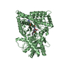
| Title | Crystal structure of a glycoside hydrolase family 3 beta-glucosidase, Bgl1 from Hypocrea jecorina at 2.1A resolution. |
|---|
 Components Components | BETA-D-GLUCOSIDE GLUCOHYDROLASE |
|---|
 Keywords Keywords |  HYDROLASE HYDROLASE |
|---|
| Function / homology |  Function and homology information Function and homology information
glucan catabolic process / scopolin beta-glucosidase activity / beta-glucosidase / beta-glucosidase activity / extracellular regionSimilarity search - Function Glycoside hydrolase family 3 C-terminal domain / Fibronectin type III-like domain / Fibronectin type III-like domain / Fibronectin type III-like domain / Glycoside hydrolase, family 3, N-terminal domain / Glycoside hydrolase family 3 C-terminal domain / Glycosyl hydrolase family 3 C-terminal domain / Glycoside hydrolase family 3 C-terminal domain superfamily / Glycoside hydrolase, family 3, N-terminal / Glycoside hydrolase, family 3, N-terminal domain superfamily ...Glycoside hydrolase family 3 C-terminal domain / Fibronectin type III-like domain / Fibronectin type III-like domain / Fibronectin type III-like domain / Glycoside hydrolase, family 3, N-terminal domain / Glycoside hydrolase family 3 C-terminal domain / Glycosyl hydrolase family 3 C-terminal domain / Glycoside hydrolase family 3 C-terminal domain superfamily / Glycoside hydrolase, family 3, N-terminal / Glycoside hydrolase, family 3, N-terminal domain superfamily / Glycosyl hydrolase family 3 N terminal domain / Glycoside hydrolase superfamily / Immunoglobulins / TIM Barrel / Alpha-Beta Barrel / Immunoglobulin-like fold / Immunoglobulin-like / Sandwich / Rossmann fold / 3-Layer(aba) Sandwich / Mainly Beta / Alpha BetaSimilarity search - Domain/homology |
|---|
| Biological species |  HYPOCREA JECORINA (fungus) HYPOCREA JECORINA (fungus) |
|---|
| Method | X-RAY DIFFRACTION / SYNCHROTRON / MOLECULAR REPLACEMENT / Resolution: 2.1 Å |
|---|
 Authors Authors | Sandgren, M. / Kaper, T. / Mikkelsen, N.E. / Hansson, H. / Piens, K. / Gudmundsson, M. / Larenas, E. / Kelemen, B. / Karkehabadi, S. |
|---|
 Citation Citation | Journal: J.Biol.Chem. / Year: 2014
Title: Biochemical Characterization and Crystal Structures of a Fungal Family 3 Beta-Glucosidase, Cel3A from Hypocrea Jecorina.
Authors: Karkehabadi, S. / Helmich, K.E. / Kaper, T. / Hansson, H. / Mikkelsen, N.E. / Gudmundsson, M. / Piens, K. / Fujdala, M. / Banerjee, G. / Scott-Craig, J.S. / Walton, J.D. / Phillips, G.N.J. / Sandgren, M. |
|---|
| History | | Deposition | Aug 30, 2011 | Deposition site: PDBE / Processing site: PDBE |
|---|
| Revision 1.0 | Dec 12, 2012 | Provider: repository / Type: Initial release |
|---|
| Revision 1.1 | Oct 22, 2014 | Group: Database references |
|---|
| Revision 1.2 | Nov 19, 2014 | Group: Database references |
|---|
| Revision 1.3 | Jul 29, 2020 | Group: Data collection / Derived calculations ...Data collection / Derived calculations / Other / Structure summary
Category: chem_comp / entity ...chem_comp / entity / pdbx_chem_comp_identifier / pdbx_database_status / pdbx_entity_nonpoly / struct_conn / struct_site / struct_site_gen
Item: _chem_comp.name / _chem_comp.type ..._chem_comp.name / _chem_comp.type / _entity.pdbx_description / _pdbx_database_status.status_code_sf / _pdbx_entity_nonpoly.name / _struct_conn.pdbx_leaving_atom_flag / _struct_conn.pdbx_role
Description: Carbohydrate remediation / Provider: repository / Type: Remediation |
|---|
|
|---|
 Movie
Movie Controller

 Yorodumi
 Open data
 Basic information
 Structure visualization
 Downloads & links
 Other downloads


 PDBj
PDBj


 Assembly
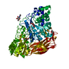


 Type: D-saccharide, beta linking / Mass: 221.208 Da / Num. of mol.: 2
 Type: D-saccharide, beta linking / Mass: 180.156 Da / Num. of mol.: 1


 Mass: 92.094 Da / Num. of mol.: 1 / Source method: obtained synthetically / Formula: C3H8O3
 Mass: 106.120 Da / Num. of mol.: 2 / Source method: obtained synthetically / Formula: C4H10O3
 Sample preparation
 / Beamline: BM14 / Wavelength: 0.95373
  Processing