 Movie
Movie Controller

[English] 日本語
 Yorodumi
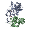
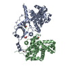
- PDB-4crl: Crystal structure of human CDK8-Cyclin C in complex with cortistatin A -
+ Open data

- Basic information
| Entry | Database: PDB / ID: 4crl | ||||||
|---|---|---|---|---|---|---|---|
| Title | Crystal structure of human CDK8-Cyclin C in complex with cortistatin A | ||||||
 Components Components |
| ||||||
 Keywords Keywords |  TRANSFERASE / CYCLIN-DEPENDENT KINASE 8 / CDK8 / CYCLIN C / CCNC / CORTISTATIN A / MEDIATOR KINASE / MEDIATOR COMPLEX / SUPER-ENHANCER / TRANSCRIPTION TRANSFERASE / CYCLIN-DEPENDENT KINASE 8 / CDK8 / CYCLIN C / CCNC / CORTISTATIN A / MEDIATOR KINASE / MEDIATOR COMPLEX / SUPER-ENHANCER / TRANSCRIPTION | ||||||
| Function / homology |  Function and homology information Function and homology informationCKM complex / G0 to G1 transition / negative regulation of triglyceride metabolic process / mediator complex / Generic Transcription Pathway / [RNA-polymerase]-subunit kinase / cyclin-dependent protein serine/threonine kinase regulator activity / negative regulation of Notch signaling pathway / cyclin-dependent protein kinase holoenzyme complex / cyclin-dependent kinase ...CKM complex / G0 to G1 transition / negative regulation of triglyceride metabolic process / mediator complex / Generic Transcription Pathway / [RNA-polymerase]-subunit kinase / cyclin-dependent protein serine/threonine kinase regulator activity / negative regulation of Notch signaling pathway / cyclin-dependent protein kinase holoenzyme complex / cyclin-dependent kinase / cyclin-dependent protein serine/threonine kinase activity / ubiquitin ligase complex / RNA polymerase II CTD heptapeptide repeat kinase activity / SMAD2/SMAD3:SMAD4 heterotrimer regulates transcription / PPARA activates gene expression / NOTCH1 Intracellular Domain Regulates Transcription / Constitutive Signaling by NOTCH1 PEST Domain Mutants / Constitutive Signaling by NOTCH1 HD+PEST Domain Mutants / Transcriptional regulation of white adipocyte differentiation / ubiquitin protein ligase activity / protein ubiquitination / protein kinase activity / phosphorylation / protein serine kinase activity / protein serine/threonine kinase activity / nucleolus / positive regulation of transcription by RNA polymerase II / protein-containing complex / nucleoplasm / ATP binding / identical protein binding / nucleusSimilarity search - Function | ||||||
| Biological species |  HOMO SAPIENS (human) HOMO SAPIENS (human) | ||||||
| Method | X-RAY DIFFRACTION / SYNCHROTRON / MOLECULAR REPLACEMENT / Resolution: 2.4 Å | ||||||
 Authors Authors | Myers, A.G. / Shair, M.D. | ||||||
 Citation Citation | Journal: Nature / Year: 2015 Title: Mediator Kinase Inhibition Further Activates Super-Enhancer- Associated Genes in Aml Authors: Pelish, H.E. / Liau, B.B. / Nitulescu, I.I. / Tangpeerachaikul, A. / Poss, Z.C. / Da Silva, D.H. / Caruso, B.T. / Arefolov, A. / Fadeyi, O. / Christie, A.L. / Du, K. / Banka, D. / Schneider, ...Authors: Pelish, H.E. / Liau, B.B. / Nitulescu, I.I. / Tangpeerachaikul, A. / Poss, Z.C. / Da Silva, D.H. / Caruso, B.T. / Arefolov, A. / Fadeyi, O. / Christie, A.L. / Du, K. / Banka, D. / Schneider, E.V. / Jestel, A. / Zou, G. / Si, C. / Ebmeier, C.C. / Bronson, R.T. / Krivtsov, A.V. / Myers, A.G. / Kohl, N.E. / Kung, A.L. / Armstrong, S.A. / Lemieux, M.E. / Taatjes, D.J. / Shair, M.D. | ||||||
| History |
|
- Structure visualization
| Structure viewer | Molecule: MolmilJmol/JSmol |
|---|
- Downloads & links
-Download
| PDBx/mmCIF format | 4crl.cif.gz | 269.4 KB | Display | PDBx/mmCIF format |
|---|---|---|---|---|
| PDB format | pdb4crl.ent.gz | 214.8 KB | Display | PDB format |
| PDBx/mmJSON format | 4crl.json.gz | Tree view | PDBx/mmJSON format | |
| Others |  Other downloads Other downloads |
-Validation report
| Arichive directory | https://data.pdbj.org/pub/pdb/validation_reports/cr/4crlftp://data.pdbj.org/pub/pdb/validation_reports/cr/4crl | HTTPS FTP |
|---|
-Related structure data
| Related structure data |  3rgfS S: Starting model for refinement |
|---|---|
| Similar structure data |


-Links
 PDBj
PDBj


- Assembly
| Deposited unit | 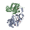
| ||||||||
|---|---|---|---|---|---|---|---|---|---|
| 1 |
| ||||||||
| Unit cell |
|
-Components
| #1: Protein | / CELL DIVISION PROTEIN KINASE 8 / MEDIATOR COMPLEX SUBUNIT CDK8 / MEDIATOR OF RNA POLYMERASE II ...CELL DIVISION PROTEIN KINASE 8 / MEDIATOR COMPLEX SUBUNIT CDK8 / MEDIATOR OF RNA POLYMERASE II TRANSCRIPTION SUBUNIT CDK8 / PROTEIN KINASE K35 / CDK8 Mass: 47105.887 Da / Num. of mol.: 1 / Fragment: KINASE DOMAIN, RESIDUES 1-403 Source method: isolated from a genetically manipulated source Source: (gene. exp.) HOMO SAPIENS (human) / Production host:   SPODOPTERA FRUGIPERDA (fall armyworm) / References: UniProt: P49336, cyclin-dependent kinase SPODOPTERA FRUGIPERDA (fall armyworm) / References: UniProt: P49336, cyclin-dependent kinase | ||
|---|---|---|---|
| #2: Protein | Mass: 33479.953 Da / Num. of mol.: 1 Source method: isolated from a genetically manipulated source Source: (gene. exp.) HOMO SAPIENS (human) / Production host: SPODOPTERA FRUGIPERDA (fall armyworm) / References: UniProt: P24863 | ||
| #3: Chemical | ChemComp-C1I / Cortistatins  Mass: 472.618 Da / Num. of mol.: 1 / Source method: obtained synthetically / Formula: C30H36N2O3 / Comment: inhibitor, alkaloid*YM Mass: 472.618 Da / Num. of mol.: 1 / Source method: obtained synthetically / Formula: C30H36N2O3 / Comment: inhibitor, alkaloid*YM | ||
| #4: Chemical | ChemComp-FMT / Formic acid  Mass: 46.025 Da / Num. of mol.: 5 / Source method: obtained synthetically / Formula: CH2O2 Mass: 46.025 Da / Num. of mol.: 5 / Source method: obtained synthetically / Formula: CH2O2#5: Water | ChemComp-HOH / | Water Mass: 18.015 Da / Num. of mol.: 103 / Source method: isolated from a natural source / Formula: H2O Mass: 18.015 Da / Num. of mol.: 103 / Source method: isolated from a natural source / Formula: H2O |
-Experimental details
-Experiment
| Experiment | Method: X-RAY DIFFRACTION / Number of used crystals: 1 |
|---|
- Sample preparation
| Crystal | Density Matthews: 3.04 Å3/Da / Density % sol: 59.61 % / Description: NONE |
|---|---|
| Crystal grow | Temperature: 293 K / Method: vapor diffusion, hanging drop Details: 20% PEG3350, 0.2M LITHIUM CHLORIDE, VAPOR DIFFUSION, HANGING DROP, TEMPERATURE 293K |
-Data collection
| Diffraction | Mean temperature: 100 K |
|---|---|
| Diffraction source | Source: SYNCHROTRON / Site: SLS  / Beamline: X06SA / Wavelength: 1.00004 / Beamline: X06SA / Wavelength: 1.00004 |
| Detector | Type: DECTRIS PILATUS 6M / Detector: PIXEL / Date: Jul 24, 2012 |
| Radiation | Protocol: SINGLE WAVELENGTH / Monochromatic (M) / Laue (L): M / Scattering type: x-ray |
| Radiation wavelength | Wavelength: 1.00004 Å / Relative weight: 1 |
| Reflection | Resolution: 2.4→85.62 Å / Num. obs: 34548 / % possible obs: 94.9 % / Observed criterion σ(I): -3 / Redundancy: 2.8 % / Rmerge(I) obs: 0.07 |
| Reflection shell | Resolution: 2.4→2.65 Å / Redundancy: 2.8 % / Rmerge(I) obs: 0.45 / % possible all: 98.6 |
- Processing
| Software |
| ||||||||||||||||||||||||||||||||||||||||||||||||||||||||||||||||||||||||||||||||||||||||||||||||||||||||||||||||||||||||||||||||||||||||||||||||||||||||||||||||||||||||||||||||||||||
|---|---|---|---|---|---|---|---|---|---|---|---|---|---|---|---|---|---|---|---|---|---|---|---|---|---|---|---|---|---|---|---|---|---|---|---|---|---|---|---|---|---|---|---|---|---|---|---|---|---|---|---|---|---|---|---|---|---|---|---|---|---|---|---|---|---|---|---|---|---|---|---|---|---|---|---|---|---|---|---|---|---|---|---|---|---|---|---|---|---|---|---|---|---|---|---|---|---|---|---|---|---|---|---|---|---|---|---|---|---|---|---|---|---|---|---|---|---|---|---|---|---|---|---|---|---|---|---|---|---|---|---|---|---|---|---|---|---|---|---|---|---|---|---|---|---|---|---|---|---|---|---|---|---|---|---|---|---|---|---|---|---|---|---|---|---|---|---|---|---|---|---|---|---|---|---|---|---|---|---|---|---|---|---|
| Refinement | Method to determine structure: MOLECULAR REPLACEMENT Starting model: PDB ENTRY 3RGF Resolution: 2.4→85.62 Å / Cor.coef. Fo:Fc: 0.945 / Cor.coef. Fo:Fc free: 0.911 / SU B: 18.294 / SU ML: 0.195 / Cross valid method: THROUGHOUT / ESU R: 0.325 / ESU R Free: 0.252 / Stereochemistry target values: MAXIMUM LIKELIHOOD Details: U VALUES WITH TLS ADDED. HYDROGENS HAVE BEEN ADDED IN THE RIDING POSITIONS
| ||||||||||||||||||||||||||||||||||||||||||||||||||||||||||||||||||||||||||||||||||||||||||||||||||||||||||||||||||||||||||||||||||||||||||||||||||||||||||||||||||||||||||||||||||||||
| Solvent computation | Ion probe radii: 0.8 Å / Shrinkage radii: 0.8 Å / VDW probe radii: 1.2 Å / Solvent model: MASK | ||||||||||||||||||||||||||||||||||||||||||||||||||||||||||||||||||||||||||||||||||||||||||||||||||||||||||||||||||||||||||||||||||||||||||||||||||||||||||||||||||||||||||||||||||||||
| Displacement parameters | Biso mean: 31.69 Å2
| ||||||||||||||||||||||||||||||||||||||||||||||||||||||||||||||||||||||||||||||||||||||||||||||||||||||||||||||||||||||||||||||||||||||||||||||||||||||||||||||||||||||||||||||||||||||
| Refinement step | Cycle: LAST / Resolution: 2.4→85.62 Å
| ||||||||||||||||||||||||||||||||||||||||||||||||||||||||||||||||||||||||||||||||||||||||||||||||||||||||||||||||||||||||||||||||||||||||||||||||||||||||||||||||||||||||||||||||||||||
| Refine LS restraints |
|