 Movie
Movie Controller

[English] 日本語
 Yorodumi
- PDB-4cla: ALTERNATIVE BINDING MODES FOR CHLORAMPHENICOL AND 1-SUBSTITUTED C... -
+ Open data

- Basic information
| Entry | Database: PDB / ID: 4cla | ||||||
|---|---|---|---|---|---|---|---|
| Title | ALTERNATIVE BINDING MODES FOR CHLORAMPHENICOL AND 1-SUBSTITUTED CHLORAMPHENICOL ANALOGUES REVEALED BY SITE-DIRECTED MUTAGENESIS AND X-RAY CRYSTALLOGRAPHY OF CHLORAMPHENICOL ACETYLTRANSFERASE | ||||||
 Components Components | TYPE III CHLORAMPHENICOL ACETYLTRANSFERASE | ||||||
 Keywords Keywords | TRANSFERASE (ACYLTRANSFERASE) | ||||||
| Function / homology |  Function and homology information Function and homology information chloramphenicol O-acetyltransferase activity / chloramphenicol O-acetyltransferase / response to antibiotic chloramphenicol O-acetyltransferase activity / chloramphenicol O-acetyltransferase / response to antibioticSimilarity search - Function | ||||||
| Biological species |  Escherichia coli (E. coli) Escherichia coli (E. coli) | ||||||
| Method | X-RAY DIFFRACTION / Resolution: 2 Å | ||||||
 Authors Authors | Leslie, A.G.W. | ||||||
 Citation Citation | Journal: Biochemistry / Year: 1991 Title: Alternative binding modes for chloramphenicol and 1-substituted chloramphenicol analogues revealed by site-directed mutagenesis and X-ray crystallography of chloramphenicol acetyltransferase. Authors: Murray, I.A. / Lewendon, A. / Williams, J.A. / Cullis, P.M. / Shaw, W.V. #1: Journal: Annu.Rev.Biophys.Biophys.Chem. / Year: 1991Title: Chloramphenicol Acetyltransferase Authors: Shaw, W.V. / Leslie, A.G.W. #2: Journal: J.Mol.Biol. / Year: 1990Title: Refined Crystal Structure of Type III Chloramphenicol Acetyltransferase at 1.75 Angstroms Resolution Authors: Leslie, A.G.W. #3: Journal: Biochemistry / Year: 1990Title: Crystal Structure of the Asp-199-Asn Mutant of Chloramphenicol Acetyltransferase to 2.35 Angstroms Resolution. Structural Consequences of Disruption of a Buried Salt-Bridge. Authors: Gibbs, M.R. / Moody, P.C.E. / Leslie, A.G.W. #4: Journal: Biochemistry / Year: 1990Title: Evidence for Transition-State Stabilization by Serine-148 in the Catalytic Mechanism of Chloramphenicol Acetyltransferase Authors: Lewendon, A. / Murray, I.A. / Shaw, W.V. / Gibbs, M.R. / Leslie, A.G.W. #5: Journal: Biochemistry / Year: 1988Title: Substitutions in the Active Site of Chloramphenicol Acetyltransferase. Role of a Conserved Aspartate Authors: Lewendon, A. / Murray, I.A. / Kleanthous, C. / Cullis, P.M. / Shaw, W.V. #6: Journal: Proc.Natl.Acad.Sci.USA / Year: 1988Title: Structure of Chloramphenicol Acetyltransferase at 1.75-Angstroms Resolution Authors: Leslie, A.G.W. / Moody, P.C.E. / Shaw, W.V. #7: Journal: J.Mol.Biol. / Year: 1986Title: Crystallization of a Type III Chloramphenicol Acetyl Transferase Authors: Leslie, A.G.W. / Liddell, J.M. / Shaw, W.V. | ||||||
| History |
| ||||||
| Remark 700 | SHEET SHEET 1 ACTUALLY HAS SEVEN STRANDS. RESIDUES 157 - 162 FORM AN EXTENSION TO THE SIX STRANDED ...SHEET SHEET 1 ACTUALLY HAS SEVEN STRANDS. RESIDUES 157 - 162 FORM AN EXTENSION TO THE SIX STRANDED BETA- SHEET OF AN ADJACENT SUBUNIT OF THE TRIMER, RESULTING IN A SEVEN-STRANDED SHEET WHICH SPANS THE SUBUNIT INTERFACE. THE FINAL STRAND, SER 157 - VAL 162, IS IN AN ADJACENT (THREE-FOLD RELATED) SUBUNIT OF THE TRIMER. N ASN 159 IS HYDROGEN BONDED TO O SEH 34. THERE IS A WIDE BETA-BULGE INVOLVING RESIDUES LYS 177, TYR 178, AND LEU 187. |
- Structure visualization
| Structure viewer | Molecule: MolmilJmol/JSmol |
|---|
- Downloads & links
-Download
| PDBx/mmCIF format | 4cla.cif.gz | 64.1 KB | Display | PDBx/mmCIF format |
|---|---|---|---|---|
| PDB format | pdb4cla.ent.gz | 46.3 KB | Display | PDB format |
| PDBx/mmJSON format | 4cla.json.gz | Tree view | PDBx/mmJSON format | |
| Others |  Other downloads Other downloads |
-Validation report
| Arichive directory | https://data.pdbj.org/pub/pdb/validation_reports/cl/4claftp://data.pdbj.org/pub/pdb/validation_reports/cl/4cla | HTTPS FTP |
|---|
-Related structure data
| Similar structure data |
|---|


-Links
 PDBj
PDBj


- Assembly
| Deposited unit | 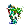
| |||||||||||||||
|---|---|---|---|---|---|---|---|---|---|---|---|---|---|---|---|---|
| 1 | 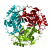
| |||||||||||||||
| 2 | x 6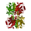
| |||||||||||||||
| Unit cell |
| |||||||||||||||
| Atom site foot note | 1: THERE IS A WIDE BETA-BULGE INVOLVING RESIDUES LYS 177, TYR 178, AND LEU 187. 2: RESIDUES 157 - 162 FORM AN EXTENSION TO THE SIX STRANDED BETA-SHEET OF AN ADJACENT SUBUNIT OF THE TRIMER, RESULTING IN A SEVEN-STRANDED SHEET WHICH SPANS THE SUBUNIT INTERFACE. | |||||||||||||||
| Components on special symmetry positions |
| |||||||||||||||
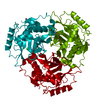
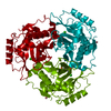
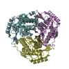
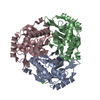
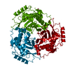
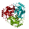
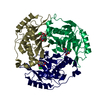
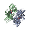
| Details | CAT IS A TRIMER OF IDENTICAL SUBUNITS EACH CONTAINING 213 AMINO ACIDS (SUBUNIT MOLECULAR WEIGHT 25000). THE THREE-FOLD AXIS OF THE TRIMER IS COINCIDENT WITH THE CRYSTALLOGRAPHIC THREE-FOLD AXIS. THE COORDINATES PRESENTED IN THIS ENTRY ARE FOR ONE SUBUNIT OF THE TRIMER. THE COORDINATES FOR THE OTHER TWO SUBUNITS CAN BE DERIVED BY APPLYING ROTATIONS OF 120 DEGREES AND 240 DEGREES ABOUT THE Z AXIS. TO GENERATE THESE SYMMETRY RELATED SUBUNITS APPLY THE FOLLOWING MATRICES TO THE COORDINATES GIVEN BELOW: 1. -0.50000 -0.86603 0.00000 0.86603 -0.50000 0.00000 0.00000 0.00000 1.00000 2. -0.50000 0.86603 0.00000 -0.86603 -0.50000 0.00000 0.00000 0.00000 1.00000 |
-Components
| #1: Protein | Mass: 25055.510 Da / Num. of mol.: 1 Source method: isolated from a genetically manipulated source Source: (gene. exp.) Escherichia coli (E. coli)References: UniProt: P00484, chloramphenicol O-acetyltransferase | ||||||||||
|---|---|---|---|---|---|---|---|---|---|---|---|
| #2: Chemical |   Mass: 58.933 Da / Num. of mol.: 2 / Source method: obtained synthetically / Formula: Co Mass: 58.933 Da / Num. of mol.: 2 / Source method: obtained synthetically / Formula: Co#3: Chemical | ChemComp-CLM / | Chloramphenicol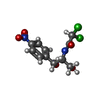  Mass: 323.129 Da / Num. of mol.: 1 / Source method: obtained synthetically / Formula: C11H12Cl2N2O5 / Comment: antibiotic*YM Mass: 323.129 Da / Num. of mol.: 1 / Source method: obtained synthetically / Formula: C11H12Cl2N2O5 / Comment: antibiotic*YM#4: Water | ChemComp-HOH / | Water Mass: 18.015 Da / Num. of mol.: 203 / Source method: isolated from a natural source / Formula: H2O Mass: 18.015 Da / Num. of mol.: 203 / Source method: isolated from a natural source / Formula: H2OCompound details | THE MUTATED RESIDUE FORMS PART OF THE CHLORAMPHENICOL BINDING POCKET. THE CONFORMATION OF THE ...THE MUTATED RESIDUE FORMS PART OF THE CHLORAMPHE | Nonpolymer details | SOLVENT ATOMS (COBALT OR WATER) LYING ON CRYSTALLOGRAPHIC SYMMETRY AXES HAVE BEEN GIVEN OCCUPANCIES ...SOLVENT ATOMS (COBALT OR WATER) LYING ON CRYSTALLOG | Sequence details | THE NUMBERING SCHEME ADOPTED IS BASED ON THE ALIGNMENT OF A NUMBER OF CAT SEQUENCES. FOR THE TYPE ...THE NUMBERING SCHEME ADOPTED IS BASED ON THE ALIGNMENT OF A NUMBER OF CAT SEQUENCES. FOR THE TYPE III ENZYME WHOSE COORDINATE | |
-Experimental details
-Experiment
| Experiment | Method: X-RAY DIFFRACTION |
|---|
- Sample preparation
| Crystal | Density Matthews: 2.76 Å3/Da / Density % sol: 55.46 % | |||||||||||||||||||||||||||||||||||
|---|---|---|---|---|---|---|---|---|---|---|---|---|---|---|---|---|---|---|---|---|---|---|---|---|---|---|---|---|---|---|---|---|---|---|---|---|
| Crystal grow | *PLUS Temperature: 4 ℃ / Method: microdialysis / pH: 6.3 | |||||||||||||||||||||||||||||||||||
| Components of the solutions | *PLUS
|
-Data collection
| Reflection | *PLUS Highest resolution: 2 Å |
|---|
- Processing
| Software | Name: DERIV / Classification: refinement | ||||||||||||||||||||||||||||||||||||||||||||||||||||||||||||
|---|---|---|---|---|---|---|---|---|---|---|---|---|---|---|---|---|---|---|---|---|---|---|---|---|---|---|---|---|---|---|---|---|---|---|---|---|---|---|---|---|---|---|---|---|---|---|---|---|---|---|---|---|---|---|---|---|---|---|---|---|---|
| Refinement | Resolution: 2→6 Å / Rfactor Rwork: 0.157 Details: ATOMS OF SIDECHAINS SHOWING VERY WEAK (LESS THAN 0.2 $E/A**3) OR UNINTERPRETABLE ELECTRON DENSITY HAVE BEEN OMITTED FROM THE MODEL. | ||||||||||||||||||||||||||||||||||||||||||||||||||||||||||||
| Refinement step | Cycle: LAST / Resolution: 2→6 Å
| ||||||||||||||||||||||||||||||||||||||||||||||||||||||||||||
| Refine LS restraints |
| ||||||||||||||||||||||||||||||||||||||||||||||||||||||||||||
| Software | *PLUS Name: PROLSQ / Classification: refinement | ||||||||||||||||||||||||||||||||||||||||||||||||||||||||||||
| Refinement | *PLUS Highest resolution: 2 Å / Lowest resolution: 6 Å / Num. reflection all: 17970 / Rfactor obs: 0.147 | ||||||||||||||||||||||||||||||||||||||||||||||||||||||||||||
| Solvent computation | *PLUS | ||||||||||||||||||||||||||||||||||||||||||||||||||||||||||||
| Displacement parameters | *PLUS | ||||||||||||||||||||||||||||||||||||||||||||||||||||||||||||
| Refine LS restraints | *PLUS
|
