 Movie
Movie Controller

[English] 日本語
 Yorodumi
- PDB-2cla: CRYSTAL STRUCTURE OF THE ASP-199-ASN MUTANT OF CHLORAMPHENICOL AC... -
+ Open data

- Basic information
| Entry | Database: PDB / ID: 2cla | ||||||
|---|---|---|---|---|---|---|---|
| Title | CRYSTAL STRUCTURE OF THE ASP-199-ASN MUTANT OF CHLORAMPHENICOL ACETYLTRANSFERASE TO 2.35 ANGSTROMS RESOLUTION. STRUCTURAL CONSEQUENCES OF DISRUPTION OF A BURIED SALT-BRIDGE | ||||||
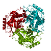
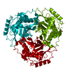
 Components Components | CHLORAMPHENICOL ACETYLTRANSFERASE | ||||||
 Keywords Keywords | TRANSFERASE (ACYLTRANSFERASE) | ||||||
| Function / homology |  Function and homology informationchloramphenicol O-acetyltransferase / chloramphenicol O-acetyltransferase activity / response to antibiotic Function and homology informationchloramphenicol O-acetyltransferase / chloramphenicol O-acetyltransferase activity / response to antibioticSimilarity search - Function | ||||||
| Biological species |  Escherichia coli (E. coli) Escherichia coli (E. coli) | ||||||
| Method | X-RAY DIFFRACTION / Resolution: 2.35 Å | ||||||
 Authors Authors | Gibbs, M.R. / Moody, P.C.E. / Leslie, A.G.W. | ||||||
 Citation Citation | Journal: Biochemistry / Year: 1990 Title: Crystal structure of the aspartic acid-199----asparagine mutant of chloramphenicol acetyltransferase to 2.35-A resolution: structural consequences of disruption of a buried salt bridge. Authors: Gibbs, M.R. / Moody, P.C. / Leslie, A.G. #1: Journal: Biochemistry / Year: 1990Title: Evidence for Transition-State Stabilization by Serine-148 in the Catalytic Mechanism of Chloramphenicol Acetyltransferase Authors: Lewendon, A. / Murray, I.A. / Shaw, W.V. / Gibbs, M.R. / Leslie, A.G.W. #2: Journal: J.Mol.Biol. / Year: 1990Title: Refined Crystal Structure of Type III Chloramphenicol Acetyltransferase at 1.75 Angstroms Resolution Authors: Leslie, A.G.W. #3: Journal: Biochemistry / Year: 1988Title: Substitutions in the Active Site of Chloramphenicol Acetyltransferase. Role of a Conserved Aspartate Authors: Lewendon, A. / Murray, I.A. / Kleanthous, C. / Cullis, P.M. / Shaw, W.V. #4: Journal: Proc.Natl.Acad.Sci.USA / Year: 1988Title: Structure of Chloramphenicol Acetyltransferase at 1.75-Angstroms Resolution Authors: Leslie, A.G.W. / Moody, P.C.E. / Shaw, W.V. #5: Journal: J.Mol.Biol. / Year: 1986Title: Crystallization of a Type III Chloramphenicol Acetyl Transferase Authors: Leslie, A.G.W. / Liddell, J.M. / Shaw, W.V. | ||||||
| History |
| ||||||
| Remark 700 | SHEET SHEET 1 ACTUALLY HAS SEVEN STRANDS. DATA BANK CONVENTIONS DO NOT ALLOW LISTING RESIDUES WHICH ...SHEET SHEET 1 ACTUALLY HAS SEVEN STRANDS. DATA BANK CONVENTIONS DO NOT ALLOW LISTING RESIDUES WHICH ARE SYMMETRY RELATED. THE FINAL STRAND, SER 157 - VAL 162, IS IN AN ADJACENT (THREE-FOLD RELATED) SUBUNIT OF THE TRIMER. N OF ASN 159 IS HYDROGEN BONDED TO O OF SEH 34. THERE IS A WIDE BETA-BULGE INVOLVING RESIDUES LYS 177, TYR 178, AND LEU 187. RESIDUES 157 - 162 FORM AN EXTENSION TO THE SIX STRANDED BETA-SHEET OF AN ADJACENT SHEET WHICH SPANS THE SUBUNIT INTERFACE. |
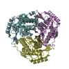
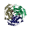
- Structure visualization
| Structure viewer | Molecule: MolmilJmol/JSmol |
|---|
- Downloads & links
-Download
| PDBx/mmCIF format | 2cla.cif.gz | 58.5 KB | Display | PDBx/mmCIF format |
|---|---|---|---|---|
| PDB format | pdb2cla.ent.gz | 41.6 KB | Display | PDB format |
| PDBx/mmJSON format | 2cla.json.gz | Tree view | PDBx/mmJSON format | |
| Others |  Other downloads Other downloads |
-Validation report
| Arichive directory | https://data.pdbj.org/pub/pdb/validation_reports/cl/2claftp://data.pdbj.org/pub/pdb/validation_reports/cl/2cla | HTTPS FTP |
|---|
-Related structure data
| Similar structure data |
|---|


-Links
 PDBj
PDBj


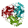
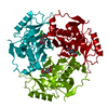
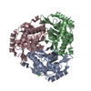
- Assembly
| Deposited unit | 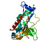
| ||||||||||||||||||
|---|---|---|---|---|---|---|---|---|---|---|---|---|---|---|---|---|---|---|---|
| 1 | 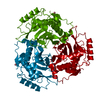
| ||||||||||||||||||
| 2 | x 6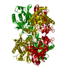
| ||||||||||||||||||
| Unit cell |
| ||||||||||||||||||
| Atom site foot note | 1: THERE IS A WIDE BETA-BULGE INVOLVING RESIDUES LYS 177, TYR 178, AND LEU 187. 2: RESIDUES 157 - 162 FORM AN EXTENSION TO THE SIX STRANDED BETA-SHEET OF AN ADJACENT SHEET WHICH SPANS THE SUBUNIT INTERFACE. | ||||||||||||||||||
| Components on special symmetry positions |
|
-Components
| #1: Protein | Mass: 25020.508 Da / Num. of mol.: 1 Source method: isolated from a genetically manipulated source Source: (gene. exp.) Escherichia coli (E. coli)References: UniProt: P00484, chloramphenicol O-acetyltransferase | ||||||
|---|---|---|---|---|---|---|---|
| #2: Chemical |   Mass: 58.933 Da / Num. of mol.: 2 / Source method: obtained synthetically / Formula: Co Mass: 58.933 Da / Num. of mol.: 2 / Source method: obtained synthetically / Formula: Co#3: Water | ChemComp-HOH / | Water Mass: 18.015 Da / Num. of mol.: 104 / Source method: isolated from a natural source / Formula: H2O Mass: 18.015 Da / Num. of mol.: 104 / Source method: isolated from a natural source / Formula: H2OCompound details | THE MUTATED ASPARTATE RESIDUE IS BELIEVED TO HAVE A STRUCTURAL ROLE AND IS SITUATED NEAR THE ACTIVE ...THE MUTATED ASPARTATE RESIDUE IS BELIEVED TO HAVE A STRUCTURAL | Sequence details | THE NUMBERING SCHEME ADOPTED IS BASED ON THE ALIGNMENT OF A NUMBER OF CAT SEQUENCES. FOR THE TYPE ...THE NUMBERING SCHEME ADOPTED IS BASED ON THE ALIGNMENT OF A NUMBER OF CAT SEQUENCES. FOR THE TYPE III ENZYME WHOSE COORDINATE | |
-Experimental details
-Experiment
| Experiment | Method: X-RAY DIFFRACTION |
|---|
- Sample preparation
| Crystal | Density Matthews: 2.77 Å3/Da / Density % sol: 55.65 % | ||||||||||||||||||||||||||||||||||||||||
|---|---|---|---|---|---|---|---|---|---|---|---|---|---|---|---|---|---|---|---|---|---|---|---|---|---|---|---|---|---|---|---|---|---|---|---|---|---|---|---|---|---|
| Crystal grow | *PLUS Temperature: 4 ℃ / pH: 6.3 / Method: microdialysis | ||||||||||||||||||||||||||||||||||||||||
| Components of the solutions | *PLUS
|
-Data collection
| Reflection | *PLUS Highest resolution: 2.35 Å / % possible obs: 75 % / Rmerge(I) obs: 0.078 |
|---|
- Processing
| Software | Name: PROLSQ / Classification: refinement | ||||||||||||||||||||||||||||||||||||||||||||||||||||||||||||||||||||||||||||||||||||
|---|---|---|---|---|---|---|---|---|---|---|---|---|---|---|---|---|---|---|---|---|---|---|---|---|---|---|---|---|---|---|---|---|---|---|---|---|---|---|---|---|---|---|---|---|---|---|---|---|---|---|---|---|---|---|---|---|---|---|---|---|---|---|---|---|---|---|---|---|---|---|---|---|---|---|---|---|---|---|---|---|---|---|---|---|---|
| Refinement | Rfactor obs: 0.152 / Highest resolution: 2.35 Å Details: THE STRUCTURE WAS SOLVED BY MOLECULAR REPLACEMENT USING THE REFINED 1.75 ANGSTROMS RESOLUTION STRUCTURE OF THE WILD TYPE ENZYME AS A MODEL. | ||||||||||||||||||||||||||||||||||||||||||||||||||||||||||||||||||||||||||||||||||||
| Refinement step | Cycle: LAST / Highest resolution: 2.35 Å
| ||||||||||||||||||||||||||||||||||||||||||||||||||||||||||||||||||||||||||||||||||||
| Refine LS restraints |
| ||||||||||||||||||||||||||||||||||||||||||||||||||||||||||||||||||||||||||||||||||||
| Refinement | *PLUS Highest resolution: 2.35 Å / Rfactor obs: 0.152 | ||||||||||||||||||||||||||||||||||||||||||||||||||||||||||||||||||||||||||||||||||||
| Solvent computation | *PLUS | ||||||||||||||||||||||||||||||||||||||||||||||||||||||||||||||||||||||||||||||||||||
| Displacement parameters | *PLUS |
