 Movie
Movie Controller

[English] 日本語
 Yorodumi
- PDB-4cl0: Structure of the Mycobacterium tuberculosis Type II Dehydroquinas... -
+ Open data

- Basic information
| Entry | Database: PDB / ID: 4cl0 | ||||||
|---|---|---|---|---|---|---|---|
| Title | Structure of the Mycobacterium tuberculosis Type II Dehydroquinase inhibited by a 3-dehydroquinic acid derivative | ||||||
 Components Components | 3-DEHYDROQUINATE DEHYDRATASE | ||||||
 Keywords Keywords | LYASE / BACTERIAL PROTEINS / TYPE 2 DEHYDROQUINASE / INHIBITOR / PROTEIN BINDING / SHIKIMIS ACID PATHWAY / SUBSTRATE SPECIFICITY | ||||||
| Function / homology |  Function and homology information Function and homology informationquinate catabolic process / Chorismate via Shikimate Pathway / 3-dehydroquinate dehydratase / 3-dehydroquinate dehydratase activity / chorismate biosynthetic process / aromatic amino acid family biosynthetic process / amino acid biosynthetic process / cytosolSimilarity search - Function | ||||||
| Biological species |   MYCOBACTERIUM TUBERCULOSIS (bacteria) MYCOBACTERIUM TUBERCULOSIS (bacteria) | ||||||
| Method | X-RAY DIFFRACTION / SYNCHROTRON / MOLECULAR REPLACEMENT / Resolution: 3.1 Å | ||||||
 Authors Authors | Otero, J.M. / Llamas-Saiz, A.L. / Maneiro, M. / Peon, A. / Sedes, A. / Lamb, H. / Hawkins, A.R. / Gonzalez-Bello, C. / van Raaij, M.J. | ||||||
 Citation Citation | Journal: To be Published Title: Investigation of the Dehydratation Mechanism Catalyzed by the Type II Dehydroquinase Authors: Maneiro, M. / Otero, J.M. / Peon, A. / Sedes, A. / Llamas-Saiz, A.L. / Lamb, H. / Hawkins, A.R. / van Raaij, M.J. / Gonzalez-Bello, C. | ||||||
| History |
| ||||||
| Remark 650 | HELIX DETERMINATION METHOD: AUTHOR PROVIDED. | ||||||
| Remark 700 | SHEET DETERMINATION METHOD: AUTHOR PROVIDED. |
- Structure visualization
| Structure viewer | Molecule: MolmilJmol/JSmol |
|---|
- Downloads & links
-Download
| PDBx/mmCIF format | 4cl0.cif.gz | 40.6 KB | Display | PDBx/mmCIF format |
|---|---|---|---|---|
| PDB format | pdb4cl0.ent.gz | 28.8 KB | Display | PDB format |
| PDBx/mmJSON format | 4cl0.json.gz | Tree view | PDBx/mmJSON format | |
| Others |  Other downloads Other downloads |
-Validation report
| Arichive directory | https://data.pdbj.org/pub/pdb/validation_reports/cl/4cl0ftp://data.pdbj.org/pub/pdb/validation_reports/cl/4cl0 | HTTPS FTP |
|---|
-Related structure data
| Related structure data |  4ckwC  4ckxC  4ckyC 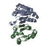 4ckzC  4v0sC  2y71S C: citing same article ( S: Starting model for refinement |
|---|---|
| Similar structure data |


-Links
 PDBj
PDBj
- Assembly
| Deposited unit | 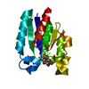
| ||||||||
|---|---|---|---|---|---|---|---|---|---|
| 1 | x 12
| ||||||||
| Unit cell |
| ||||||||
| Components on special symmetry positions |
|
-Components
| #1: Protein | / 3-DEHYDROQUINASE / TYPE II DHQASE Mass: 15676.737 Da / Num. of mol.: 1 Source method: isolated from a genetically manipulated source Source: (gene. exp.) MYCOBACTERIUM TUBERCULOSIS (bacteria) / Plasmid: PKK233-2 / Production host: ESCHERICHIA COLI (E. coli) / Strain (production host): BL21 / Variant (production host): SK3430References: UniProt: P0A4Z6, UniProt: P9WPX7*PLUS, 3-dehydroquinate dehydratase |
|---|---|
| #2: Chemical | ChemComp-9PY / (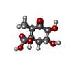  Mass: 204.177 Da / Num. of mol.: 1 / Source method: obtained synthetically / Formula: C8H12O6 Mass: 204.177 Da / Num. of mol.: 1 / Source method: obtained synthetically / Formula: C8H12O6 |
| #3: Water | ChemComp-HOH / Water Mass: 18.015 Da / Num. of mol.: 13 / Source method: isolated from a natural source / Formula: H2O Mass: 18.015 Da / Num. of mol.: 13 / Source method: isolated from a natural source / Formula: H2O |
-Experimental details
-Experiment
| Experiment | Method: X-RAY DIFFRACTION / Number of used crystals: 1 |
|---|
- Sample preparation
| Crystal | Density Matthews: 2.8 Å3/Da / Density % sol: 55.7 % / Description: NONE |
|---|---|
| Crystal grow | pH: 7.5 Details: 32% (V/V) 2-METHYL-2,4-PENTANEDIOL, 0.3 M AMMONIUM SULFATE, 0.1 M 4-(2-HYDROXYETHYL)-PIPERAZINE- 1-ETHANESULFONIC ACID SODIUM SALT (HEPES) PH 7.5 |
-Data collection
| Diffraction | Mean temperature: 100 K |
|---|---|
| Diffraction source | Source: SYNCHROTRON / Site: ESRF  / Beamline: ID29 / Wavelength: 0.97908 / Beamline: ID29 / Wavelength: 0.97908 |
| Detector | Type: DECTRIS PILATUS 6M / Detector: PIXEL / Date: May 7, 2013 / Details: CYLINDRICAL GRAZING INCIDENCE MIRROR |
| Radiation | Monochromator: CHANNEL-CUT DOUBLE CRYSTAL MONOCHROMATOR / Protocol: SINGLE WAVELENGTH / Monochromatic (M) / Laue (L): M / Scattering type: x-ray |
| Radiation wavelength | Wavelength: 0.97908 Å / Relative weight: 1 |
| Reflection | Resolution: 3.1→44.55 Å / Num. obs: 3177 / % possible obs: 100 % / Observed criterion σ(I): -3 / Redundancy: 9.8 % / Biso Wilson estimate: 39 Å2 / Rmerge(I) obs: 0.11 / Net I/σ(I): 19.9 |
| Reflection shell | Resolution: 3.1→3.27 Å / Redundancy: 9.7 % / Rmerge(I) obs: 0.35 / Mean I/σ(I) obs: 7.6 / % possible all: 100 |
- Processing
| Software |
| ||||||||||||||||||||||||||||||||||||||||||||||||||||||||||||||||||||||||||||||||||||||||||||||||||||||||||||||||||||||||||||||||||||||||||||||||||||||||||||||||||||||||||||||||||||||
|---|---|---|---|---|---|---|---|---|---|---|---|---|---|---|---|---|---|---|---|---|---|---|---|---|---|---|---|---|---|---|---|---|---|---|---|---|---|---|---|---|---|---|---|---|---|---|---|---|---|---|---|---|---|---|---|---|---|---|---|---|---|---|---|---|---|---|---|---|---|---|---|---|---|---|---|---|---|---|---|---|---|---|---|---|---|---|---|---|---|---|---|---|---|---|---|---|---|---|---|---|---|---|---|---|---|---|---|---|---|---|---|---|---|---|---|---|---|---|---|---|---|---|---|---|---|---|---|---|---|---|---|---|---|---|---|---|---|---|---|---|---|---|---|---|---|---|---|---|---|---|---|---|---|---|---|---|---|---|---|---|---|---|---|---|---|---|---|---|---|---|---|---|---|---|---|---|---|---|---|---|---|---|---|
| Refinement | Method to determine structure: MOLECULAR REPLACEMENT Starting model: PDB ENTRY 2Y71 Resolution: 3.1→44.59 Å / Cor.coef. Fo:Fc: 0.959 / Cor.coef. Fo:Fc free: 0.862 / SU B: 15.61 / SU ML: 0.267 / Cross valid method: THROUGHOUT / ESU R Free: 0.434 / Stereochemistry target values: MAXIMUM LIKELIHOOD Details: HYDROGENS HAVE BEEN ADDED IN THE RIDING POSITIONS. U VALUES ARE REFINED INDIVIDUALLY. ARG-18 AND GLU-42 SIDE CHAINS ARE MISSING BY DISORDER IN THE STRUCTURE. INHIBITOR INCLUDED IN ENZYME ...Details: HYDROGENS HAVE BEEN ADDED IN THE RIDING POSITIONS. U VALUES ARE REFINED INDIVIDUALLY. ARG-18 AND GLU-42 SIDE CHAINS ARE MISSING BY DISORDER IN THE STRUCTURE. INHIBITOR INCLUDED IN ENZYME ACTIVE SITE BY SOAKING OF APO-CRYSTALS IN INHIBITOR SOLUTION
| ||||||||||||||||||||||||||||||||||||||||||||||||||||||||||||||||||||||||||||||||||||||||||||||||||||||||||||||||||||||||||||||||||||||||||||||||||||||||||||||||||||||||||||||||||||||
| Solvent computation | Ion probe radii: 0.8 Å / Shrinkage radii: 0.8 Å / VDW probe radii: 1.2 Å / Solvent model: MASK | ||||||||||||||||||||||||||||||||||||||||||||||||||||||||||||||||||||||||||||||||||||||||||||||||||||||||||||||||||||||||||||||||||||||||||||||||||||||||||||||||||||||||||||||||||||||
| Displacement parameters | Biso mean: 47.75 Å2
| ||||||||||||||||||||||||||||||||||||||||||||||||||||||||||||||||||||||||||||||||||||||||||||||||||||||||||||||||||||||||||||||||||||||||||||||||||||||||||||||||||||||||||||||||||||||
| Refinement step | Cycle: LAST / Resolution: 3.1→44.59 Å
| ||||||||||||||||||||||||||||||||||||||||||||||||||||||||||||||||||||||||||||||||||||||||||||||||||||||||||||||||||||||||||||||||||||||||||||||||||||||||||||||||||||||||||||||||||||||
| Refine LS restraints |
|