 Movie
Movie Controller

[English] 日本語
 Yorodumi
- PDB-4kiu: Design and structural analysis of aromatic inhibitors of type II ... -
+ Open data

- Basic information
| Entry | Database: PDB / ID: 4kiu | ||||||
|---|---|---|---|---|---|---|---|
| Title | Design and structural analysis of aromatic inhibitors of type II dehydroquinate dehydratase from Mycobacterium tuberculosis - compound 49d [5-[(3-nitrobenzyl)oxy]benzene-1,3-dicarboxylic acid] | ||||||
 Components Components | 3-dehydroquinate dehydratase | ||||||
 Keywords Keywords | Lyase/Lyase Inhibitor / dehydratase / Lyase-Lyase Inhibitor complex | ||||||
| Function / homology |  Function and homology information Function and homology informationquinate catabolic process / Chorismate via Shikimate Pathway / 3-dehydroquinate dehydratase / 3-dehydroquinate dehydratase activity / chorismate biosynthetic process / aromatic amino acid family biosynthetic process / amino acid biosynthetic process / cytosolSimilarity search - Function | ||||||
| Biological species |   Mycobacterium tuberculosis (bacteria) Mycobacterium tuberculosis (bacteria) | ||||||
| Method | X-RAY DIFFRACTION / SYNCHROTRON / MOLECULAR REPLACEMENT / Resolution: 2.4 Å | ||||||
 Authors Authors | Dias, M.V.B. / Howard, N.G. / Blundell, T.L. / Abell, C. | ||||||
 Citation Citation | Journal: Chemmedchem / Year: 2015 Title: Design and Structural Analysis of Aromatic Inhibitors of Type II Dehydroquinase from Mycobacterium tuberculosis. Authors: Howard, N.I. / Dias, M.V. / Peyrot, F. / Chen, L. / Schmidt, M.F. / Blundell, T.L. / Abell, C. | ||||||
| History |
|
- Structure visualization
| Structure viewer | Molecule: MolmilJmol/JSmol |
|---|
- Downloads & links
-Download
| PDBx/mmCIF format | 4kiu.cif.gz | 650.4 KB | Display | PDBx/mmCIF format |
|---|---|---|---|---|
| PDB format | pdb4kiu.ent.gz | 540.9 KB | Display | PDB format |
| PDBx/mmJSON format | 4kiu.json.gz | Tree view | PDBx/mmJSON format | |
| Others |  Other downloads Other downloads |
-Validation report
| Arichive directory | https://data.pdbj.org/pub/pdb/validation_reports/ki/4kiuftp://data.pdbj.org/pub/pdb/validation_reports/ki/4kiu | HTTPS FTP |
|---|
-Related structure data
| Related structure data |  4ki7C  4kijC  4kiwC  4kim C: citing same article ( |
|---|---|
| Similar structure data |


-Links
 PDBj
PDBj
- Assembly
| Deposited unit | 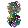
| ||||||||
|---|---|---|---|---|---|---|---|---|---|
| 1 | 
| ||||||||
| 2 | 
| ||||||||
| Unit cell |
|
-Components
| #1: Protein | / 3-dehydroquinase / Type II DHQase Mass: 16584.789 Da / Num. of mol.: 24 / Fragment: aroD Source method: isolated from a genetically manipulated source Source: (gene. exp.) Mycobacterium tuberculosis (bacteria) / Gene: aroD, aroQ, MT2612, MTCY159.19, Rv2537c / Production host: Escherichia coli (E. coli) / Strain (production host): BL21(de3)References: UniProt: P0A4Z6, UniProt: P9WPX7*PLUS, 3-dehydroquinate dehydratase#2: Chemical | ChemComp-KIU /   Mass: 317.250 Da / Num. of mol.: 22 / Source method: obtained synthetically / Formula: C15H11NO7 Mass: 317.250 Da / Num. of mol.: 22 / Source method: obtained synthetically / Formula: C15H11NO7#3: Water | ChemComp-HOH / | Water Mass: 18.015 Da / Num. of mol.: 761 / Source method: isolated from a natural source / Formula: H2O Mass: 18.015 Da / Num. of mol.: 761 / Source method: isolated from a natural source / Formula: H2O |
|---|
-Experimental details
-Experiment
| Experiment | Method: X-RAY DIFFRACTION / Number of used crystals: 1 |
|---|
- Sample preparation
| Crystal | Density Matthews: 2.4 Å3/Da / Density % sol: 48.72 % |
|---|---|
| Crystal grow | Temperature: 298 K / Method: vapor diffusion / pH: 7.8 Details: 0.15M KBr, 30% 5000mme, pH 7.8, VAPOR DIFFUSION, temperature 298K |
-Data collection
| Diffraction | Mean temperature: 100 K | ||||||||||||||||||||||||||||||||||||||||||||||||||||||||||||||||||||||||||||||||||||||||
|---|---|---|---|---|---|---|---|---|---|---|---|---|---|---|---|---|---|---|---|---|---|---|---|---|---|---|---|---|---|---|---|---|---|---|---|---|---|---|---|---|---|---|---|---|---|---|---|---|---|---|---|---|---|---|---|---|---|---|---|---|---|---|---|---|---|---|---|---|---|---|---|---|---|---|---|---|---|---|---|---|---|---|---|---|---|---|---|---|---|
| Diffraction source | Source: SYNCHROTRON / Site: ESRF  / Beamline: ID23-1 / Wavelength: 0.9791 Å / Beamline: ID23-1 / Wavelength: 0.9791 Å | ||||||||||||||||||||||||||||||||||||||||||||||||||||||||||||||||||||||||||||||||||||||||
| Detector | Type: MAR CCD 165 mm / Detector: CCD / Date: Jun 15, 2009 | ||||||||||||||||||||||||||||||||||||||||||||||||||||||||||||||||||||||||||||||||||||||||
| Radiation | Monochromator: mirrors / Protocol: SINGLE WAVELENGTH / Monochromatic (M) / Laue (L): M / Scattering type: x-ray | ||||||||||||||||||||||||||||||||||||||||||||||||||||||||||||||||||||||||||||||||||||||||
| Radiation wavelength | Wavelength: 0.9791 Å / Relative weight: 1 | ||||||||||||||||||||||||||||||||||||||||||||||||||||||||||||||||||||||||||||||||||||||||
| Reflection | Resolution: 2.4→142.722 Å / Num. all: 142809 / Num. obs: 142809 / % possible obs: 97.2 % / Observed criterion σ(F): 2 / Observed criterion σ(I): 2 / Redundancy: 2.8 % / Rsym value: 0.173 / Net I/σ(I): 5.8 | ||||||||||||||||||||||||||||||||||||||||||||||||||||||||||||||||||||||||||||||||||||||||
| Reflection shell | Diffraction-ID: 1
|
- Processing
| Software |
| |||||||||||||||||||||||||||||||||||||||||||||
|---|---|---|---|---|---|---|---|---|---|---|---|---|---|---|---|---|---|---|---|---|---|---|---|---|---|---|---|---|---|---|---|---|---|---|---|---|---|---|---|---|---|---|---|---|---|---|
| Refinement | Method to determine structure: MOLECULAR REPLACEMENT / Resolution: 2.4→45.28 Å / Cor.coef. Fo:Fc: 0.938 / Cor.coef. Fo:Fc free: 0.877 / Occupancy max: 1 / Occupancy min: 0.5 / SU B: 9.65 / SU ML: 0.22 / Cross valid method: THROUGHOUT / σ(F): 0 / ESU R: 0.497 / ESU R Free: 0.297 / Stereochemistry target values: MAXIMUM LIKELIHOOD Details: HYDROGENS HAVE BEEN USED IF PRESENT IN THE INPUT U VALUES: REFINED INDIVIDUALLY
| |||||||||||||||||||||||||||||||||||||||||||||
| Solvent computation | Ion probe radii: 0.8 Å / Shrinkage radii: 0.8 Å / VDW probe radii: 1.2 Å / Solvent model: MASK | |||||||||||||||||||||||||||||||||||||||||||||
| Displacement parameters | Biso max: 116.38 Å2 / Biso mean: 29.9535 Å2 / Biso min: 2 Å2
| |||||||||||||||||||||||||||||||||||||||||||||
| Refinement step | Cycle: LAST / Resolution: 2.4→45.28 Å
| |||||||||||||||||||||||||||||||||||||||||||||
| Refine LS restraints |
| |||||||||||||||||||||||||||||||||||||||||||||
| LS refinement shell | Resolution: 2.4→2.462 Å / Total num. of bins used: 20
|
