 Movie
Movie Controller

[English] 日本語
 Yorodumi
- PDB-4cjn: Crystal structure of PBP2a from MRSA in complex with quinazolinon... -
+ Open data

- Basic information
| Entry | Database: PDB / ID: 4cjn | ||||||
|---|---|---|---|---|---|---|---|
| Title | Crystal structure of PBP2a from MRSA in complex with quinazolinone ligand | ||||||
 Components Components | PENICILLIN BINDING PROTEIN 2 PRIME | ||||||
 Keywords Keywords |  HYDROLASE / IMMUNE SYSTEM / ALLOSTERIC SITE HYDROLASE / IMMUNE SYSTEM / ALLOSTERIC SITE | ||||||
| Function / homology |  Function and homology informationserine-type D-Ala-D-Ala carboxypeptidase / serine-type D-Ala-D-Ala carboxypeptidase activity / penicillin binding / membrane => GO:0016020 / response to antibiotic / membrane Function and homology informationserine-type D-Ala-D-Ala carboxypeptidase / serine-type D-Ala-D-Ala carboxypeptidase activity / penicillin binding / membrane => GO:0016020 / response to antibiotic / membraneSimilarity search - Function | ||||||
| Biological species |  STAPHYLOCOCCUS AUREUS SUBSP. AUREUS MU50 (bacteria) STAPHYLOCOCCUS AUREUS SUBSP. AUREUS MU50 (bacteria) | ||||||
| Method | X-RAY DIFFRACTION / SYNCHROTRON / MOLECULAR REPLACEMENT / Resolution: 1.947 Å | ||||||
 Authors Authors | Bouley, R. / Otero, L.H. / Rojas-Altuve, A. / Hermoso, J.A. | ||||||
 Citation Citation | Journal: J.Am.Chem.Soc. / Year: 2015 Title: Discovery of Antibiotic (E)-3-(3-Carboxyphenyl)-2-(4-Cyanostyryl)Quinazolin-4(3H)-One. Authors: Bouley, R. / Kumarasiri, M. / Peng, Z. / Otero, L.H. / Song, W. / Suckow, M.A. / Schroeder, V.A. / Wolter, W.R. / Lastochkin, E. / Antunes, N.T. / Pi, H. / Vakulenko, S. / Hermoso, J.A. / ...Authors: Bouley, R. / Kumarasiri, M. / Peng, Z. / Otero, L.H. / Song, W. / Suckow, M.A. / Schroeder, V.A. / Wolter, W.R. / Lastochkin, E. / Antunes, N.T. / Pi, H. / Vakulenko, S. / Hermoso, J.A. / Chang, M. / Mobashery, S. #1: Journal: Proc.Natl.Acad.Sci.USA / Year: 2013Title: How Allosteric Control of Staphylococcus Aureus Penicillin Binding Protein 2A Enables Methicillin Resistance and Physiological Function. Authors: Otero, L.H. / Rojas-Altuve, A. / Llarrull, L.I. / Carrasco-Lopez, C. / Kumarasiri, M. / Lastochkin, E. / Fishovitz, J. / Dawley, M. / Hesek, D. / Lee, M. / Johnson, J.W. / Fisher, J.F. / ...Authors: Otero, L.H. / Rojas-Altuve, A. / Llarrull, L.I. / Carrasco-Lopez, C. / Kumarasiri, M. / Lastochkin, E. / Fishovitz, J. / Dawley, M. / Hesek, D. / Lee, M. / Johnson, J.W. / Fisher, J.F. / Chang, M. / Mobashery, S. / Hermoso, J.A. | ||||||
| History |
|
- Structure visualization
| Structure viewer | Molecule: MolmilJmol/JSmol |
|---|
- Downloads & links
-Download
| PDBx/mmCIF format | 4cjn.cif.gz | 539.6 KB | Display | PDBx/mmCIF format |
|---|---|---|---|---|
| PDB format | pdb4cjn.ent.gz | 443.3 KB | Display | PDB format |
| PDBx/mmJSON format | 4cjn.json.gz | Tree view | PDBx/mmJSON format | |
| Others |  Other downloads Other downloads |
-Validation report
| Arichive directory | https://data.pdbj.org/pub/pdb/validation_reports/cj/4cjnftp://data.pdbj.org/pub/pdb/validation_reports/cj/4cjn | HTTPS FTP |
|---|
-Related structure data
| Related structure data |  3zg0S S: Starting model for refinement |
|---|---|
| Similar structure data |


-Links
 PDBj
PDBj

- Assembly
| Deposited unit | 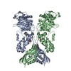
| ||||||||
|---|---|---|---|---|---|---|---|---|---|
| 1 |
| ||||||||
| Unit cell |
|
-Components
-Protein / Sugars , 2 types, 4 molecules AB
| #1: Protein | Mass: 73286.656 Da / Num. of mol.: 2 / Fragment: RESIDUES 27-668 Source method: isolated from a genetically manipulated source Source: (gene. exp.) STAPHYLOCOCCUS AUREUS SUBSP. AUREUS MU50 (bacteria)Production host: ESCHERICHIA COLI (E. coli) / Strain (production host): BL21(DE3) / Variant (production host): STARReferences: UniProt: Q54113, UniProt: A0A0J9X1X5*PLUS, serine-type D-Ala-D-Ala carboxypeptidase#4: Sugar | Muramic acid Type: D-saccharide, beta linking / Mass: 251.234 Da / Num. of mol.: 2 / Source method: obtained synthetically / Formula: C9H17NO7 Type: D-saccharide, beta linking / Mass: 251.234 Da / Num. of mol.: 2 / Source method: obtained synthetically / Formula: C9H17NO7 |
|---|
-Non-polymers , 4 types, 304 molecules 


| #2: Chemical | ChemComp-CD /  Mass: 112.411 Da / Num. of mol.: 4 / Source method: obtained synthetically / Formula: Cd Mass: 112.411 Da / Num. of mol.: 4 / Source method: obtained synthetically / Formula: Cd#3: Chemical | ChemComp-CL / Chloride Mass: 35.453 Da / Num. of mol.: 4 / Source method: obtained synthetically / Formula: Cl Mass: 35.453 Da / Num. of mol.: 4 / Source method: obtained synthetically / Formula: Cl#5: Chemical | ChemComp-QNZ / ( |  Mass: 393.394 Da / Num. of mol.: 1 / Source method: obtained synthetically / Formula: C24H15N3O3 Mass: 393.394 Da / Num. of mol.: 1 / Source method: obtained synthetically / Formula: C24H15N3O3#6: Water | ChemComp-HOH / | WaterMass: 18.015 Da / Num. of mol.: 295 / Source method: isolated from a natural source / Formula: H2O |
|---|
-Experimental details
-Experiment
| Experiment | Method: X-RAY DIFFRACTION |
|---|
- Sample preparation
| Crystal | Density Matthews: 2.67 Å3/Da / Density % sol: 54.01 % / Description: NONE |
|---|---|
| Crystal grow | pH: 7 / Details: PH 7 |
-Data collection
| Diffraction | Mean temperature: 100 K |
|---|---|
| Diffraction source | Source: SYNCHROTRON / Site: SLS  / Beamline: X06SA / Wavelength: 0.99999 / Beamline: X06SA / Wavelength: 0.99999 |
| Radiation | Protocol: SINGLE WAVELENGTH / Monochromatic (M) / Laue (L): M / Scattering type: x-ray |
| Radiation wavelength | Wavelength: 0.99999 Å / Relative weight: 1 |
| Reflection | Resolution: 1.95→63.96 Å / Num. obs: 115623 / % possible obs: 100 % / Observed criterion σ(I): 2 / Redundancy: 25.6 % / Rmerge(I) obs: 0.09 / Net I/σ(I): 19.5 |
| Reflection shell | Resolution: 1.95→2.06 Å / Redundancy: 18.4 % / Mean I/σ(I) obs: 1.9 / % possible all: 100 |
- Processing
| Software |
| |||||||||||||||||||||||||||||||||||||||||||||||||||||||||||||||||||||||||||||||||||||||||||||||||||||||||||||||||||||||||||||||||||||||||||||||||||||||||||||||||||||||||||||||||||||||||||||||||||||||||||||||||||||||||
|---|---|---|---|---|---|---|---|---|---|---|---|---|---|---|---|---|---|---|---|---|---|---|---|---|---|---|---|---|---|---|---|---|---|---|---|---|---|---|---|---|---|---|---|---|---|---|---|---|---|---|---|---|---|---|---|---|---|---|---|---|---|---|---|---|---|---|---|---|---|---|---|---|---|---|---|---|---|---|---|---|---|---|---|---|---|---|---|---|---|---|---|---|---|---|---|---|---|---|---|---|---|---|---|---|---|---|---|---|---|---|---|---|---|---|---|---|---|---|---|---|---|---|---|---|---|---|---|---|---|---|---|---|---|---|---|---|---|---|---|---|---|---|---|---|---|---|---|---|---|---|---|---|---|---|---|---|---|---|---|---|---|---|---|---|---|---|---|---|---|---|---|---|---|---|---|---|---|---|---|---|---|---|---|---|---|---|---|---|---|---|---|---|---|---|---|---|---|---|---|---|---|---|---|---|---|---|---|---|---|---|---|---|---|---|---|---|---|---|
| Refinement | Method to determine structure: MOLECULAR REPLACEMENT Starting model: PDB ENTRY 3ZG0 Resolution: 1.947→63.876 Å / SU ML: 0.26 / σ(F): 1.33 / Phase error: 33.51 / Stereochemistry target values: ML
| |||||||||||||||||||||||||||||||||||||||||||||||||||||||||||||||||||||||||||||||||||||||||||||||||||||||||||||||||||||||||||||||||||||||||||||||||||||||||||||||||||||||||||||||||||||||||||||||||||||||||||||||||||||||||
| Solvent computation | Shrinkage radii: 0.9 Å / VDW probe radii: 1.11 Å / Solvent model: FLAT BULK SOLVENT MODEL | |||||||||||||||||||||||||||||||||||||||||||||||||||||||||||||||||||||||||||||||||||||||||||||||||||||||||||||||||||||||||||||||||||||||||||||||||||||||||||||||||||||||||||||||||||||||||||||||||||||||||||||||||||||||||
| Refinement step | Cycle: LAST / Resolution: 1.947→63.876 Å
| |||||||||||||||||||||||||||||||||||||||||||||||||||||||||||||||||||||||||||||||||||||||||||||||||||||||||||||||||||||||||||||||||||||||||||||||||||||||||||||||||||||||||||||||||||||||||||||||||||||||||||||||||||||||||
| Refine LS restraints |
| |||||||||||||||||||||||||||||||||||||||||||||||||||||||||||||||||||||||||||||||||||||||||||||||||||||||||||||||||||||||||||||||||||||||||||||||||||||||||||||||||||||||||||||||||||||||||||||||||||||||||||||||||||||||||
| LS refinement shell |
| |||||||||||||||||||||||||||||||||||||||||||||||||||||||||||||||||||||||||||||||||||||||||||||||||||||||||||||||||||||||||||||||||||||||||||||||||||||||||||||||||||||||||||||||||||||||||||||||||||||||||||||||||||||||||
| Refinement TLS params. | Method: refined / Refine-ID: X-RAY DIFFRACTION
| |||||||||||||||||||||||||||||||||||||||||||||||||||||||||||||||||||||||||||||||||||||||||||||||||||||||||||||||||||||||||||||||||||||||||||||||||||||||||||||||||||||||||||||||||||||||||||||||||||||||||||||||||||||||||
| Refinement TLS group |
|
