 Movie
Movie Controller


+ Open data

- Basic information
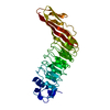
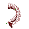
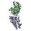
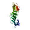
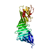
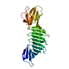
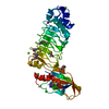
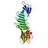
| Entry | Database: PDB / ID: 4cil | ||||||
|---|---|---|---|---|---|---|---|
| Title | YopM-InlB: Hybrid leucine-rich repeat protein | ||||||
 Components Components | YOPM-CAP, INTERNALIN B | ||||||
 Keywords Keywords |  SIGNALING PROTEIN / CAPPING / FUSION PROTEIN / LRR / PROTEIN CHIMERA / PROTEIN DESIGN / PROTEIN ENGINEERING SIGNALING PROTEIN / CAPPING / FUSION PROTEIN / LRR / PROTEIN CHIMERA / PROTEIN DESIGN / PROTEIN ENGINEERING | ||||||
| Function / homology |  Function and homology information Function and homology informationInlB-mediated entry of Listeria monocytogenes into host cell / heparin binding / lipid binding / cell surface / extracellular region / metal ion binding / plasma membraneSimilarity search - Function | ||||||
| Biological species |  YERSINIA ENTEROCOLITICA (bacteria)LISTERIA MONOCYTOGENES (bacteria) YERSINIA ENTEROCOLITICA (bacteria)LISTERIA MONOCYTOGENES (bacteria) | ||||||
| Method | X-RAY DIFFRACTION / SYNCHROTRON / MOLECULAR REPLACEMENT / Resolution: 1.5 Å | ||||||
 Authors Authors | Breitsprecher, D. / Niemann, H.H. | ||||||
 Citation Citation | Journal: Bmc Struct.Biol. / Year: 2014 Title: Crystal Structure of an Engineered Yopm-Inlb Hybrid Protein. Authors: Breitsprecher, D. / Gherardi, E. / Bleymuller, W.M. / Niemann, H.H. | ||||||
| History |
|
- Structure visualization
| Structure viewer | Molecule: MolmilJmol/JSmol |
|---|
- Downloads & links
-Download
| PDBx/mmCIF format | 4cil.cif.gz | 130.1 KB | Display | PDBx/mmCIF format |
|---|---|---|---|---|
| PDB format | pdb4cil.ent.gz | 100.9 KB | Display | PDB format |
| PDBx/mmJSON format | 4cil.json.gz | Tree view | PDBx/mmJSON format | |
| Others |  Other downloads Other downloads |
-Validation report
| Arichive directory | https://data.pdbj.org/pub/pdb/validation_reports/ci/4cilftp://data.pdbj.org/pub/pdb/validation_reports/ci/4cil | HTTPS FTP |
|---|
-Related structure data


-Links
 PDBj
PDBj


- Assembly
| Deposited unit | 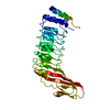
| ||||||||
|---|---|---|---|---|---|---|---|---|---|
| 1 |
| ||||||||
| Unit cell |
| ||||||||
| Components on special symmetry positions |
|
-Components
| #1: Protein | Mass: 32170.664 Da / Num. of mol.: 1 Fragment: YOPM N-TERMINAL CAP RESIDUES 34-87, INLB LEUCINE-RICH REPEAT AND INTER-REPEAT REGION RESIDUES 93-321 Source method: isolated from a genetically manipulated source Source: (gene. exp.) YERSINIA ENTEROCOLITICA (bacteria), (gene. exp.) LISTERIA MONOCYTOGENES (bacteria)Strain: W22703, EGD-E / Description: PLASMID PYVE227 / Production host: ESCHERICHIA COLI (E. coli) / Strain (production host): BL21(DE3) / Variant (production host): CODONPLUS / References: UniProt: P74988, UniProt: P25147 |
|---|---|
| #2: Water | ChemComp-HOH / Water Mass: 18.015 Da / Num. of mol.: 250 / Source method: isolated from a natural source / Formula: H2O Mass: 18.015 Da / Num. of mol.: 250 / Source method: isolated from a natural source / Formula: H2O |
| Sequence details | ONLY RESIDUES 93-321 ARE CONTAINED IN THE FUSION PROTEIN. ONLY RESIDUES 34-87 ARE CONTAINED IN THE ...ONLY RESIDUES 93-321 ARE CONTAINED IN THE FUSION PROTEIN. ONLY RESIDUES 34-87 ARE CONTAINED IN THE FUSION PROTEIN. |
-Experimental details
-Experiment
| Experiment | Method: X-RAY DIFFRACTION / Number of used crystals: 1 |
|---|
- Sample preparation
| Crystal | Density Matthews: 1.91 Å3/Da / Density % sol: 35.6 % / Description: NONE |
|---|---|
| Crystal grow | Temperature: 293 K / Method: vapor diffusion, hanging drop Details: HANGING DROP VAPOUR DIFFUSION AT 293 K WITH A DROP SIZE OF 2 UL CONSISTING OF EQUAL VOLUMES OF PROTEIN AT A CONCENTRATION OF 10 MG/ML AND RESERVOIR SOLUTION (0.1 M TRICINE, PH 9.0, 28% PEG ...Details: HANGING DROP VAPOUR DIFFUSION AT 293 K WITH A DROP SIZE OF 2 UL CONSISTING OF EQUAL VOLUMES OF PROTEIN AT A CONCENTRATION OF 10 MG/ML AND RESERVOIR SOLUTION (0.1 M TRICINE, PH 9.0, 28% PEG 1000, 10% GLYCEROL, 0.25M KCL). CRYSTALS WERE CRYO-PROTECTED WITH RESERVOIR-SOLUTION ADDITIONALLY CONTAINING 15% GLYCEROL AND FLASH-FROZEN IN LIQUID NITROGEN |
-Data collection
| Diffraction | Mean temperature: 100 K |
|---|---|
| Diffraction source | Source: SYNCHROTRON / Site: EMBL/DESY, HAMBURG  / Beamline: X13 / Wavelength: 0.81 / Beamline: X13 / Wavelength: 0.81 |
| Detector | Type: MARRESEARCH / Detector: CCD |
| Radiation | Protocol: SINGLE WAVELENGTH / Monochromatic (M) / Laue (L): M / Scattering type: x-ray |
| Radiation wavelength | Wavelength: 0.81 Å / Relative weight: 1 |
| Reflection | Resolution: 1.5→20 Å / Num. obs: 38095 / % possible obs: 96.3 % / Redundancy: 3.8 % / Rmerge(I) obs: 0.07 / Net I/σ(I): 13.88 |
| Reflection shell | Resolution: 1.5→1.54 Å / Redundancy: 3.28 % / Rmerge(I) obs: 0.82 / Mean I/σ(I) obs: 1.8 / % possible all: 82.4 |
- Processing
| Software |
| ||||||||||||||||||||||||||||||||||||||||||||||||||||||||||||||||||||||||||||||||||||||||||||||||||||||||||||||||||||||||||||||||||||||||||||||||||||||||||||||||||||||||||||||||||||||
|---|---|---|---|---|---|---|---|---|---|---|---|---|---|---|---|---|---|---|---|---|---|---|---|---|---|---|---|---|---|---|---|---|---|---|---|---|---|---|---|---|---|---|---|---|---|---|---|---|---|---|---|---|---|---|---|---|---|---|---|---|---|---|---|---|---|---|---|---|---|---|---|---|---|---|---|---|---|---|---|---|---|---|---|---|---|---|---|---|---|---|---|---|---|---|---|---|---|---|---|---|---|---|---|---|---|---|---|---|---|---|---|---|---|---|---|---|---|---|---|---|---|---|---|---|---|---|---|---|---|---|---|---|---|---|---|---|---|---|---|---|---|---|---|---|---|---|---|---|---|---|---|---|---|---|---|---|---|---|---|---|---|---|---|---|---|---|---|---|---|---|---|---|---|---|---|---|---|---|---|---|---|---|---|
| Refinement | Method to determine structure: MOLECULAR REPLACEMENT Starting model: PDB ENTRIES 1H6T AND 1JL5 Resolution: 1.5→20 Å / Cor.coef. Fo:Fc: 0.967 / Cor.coef. Fo:Fc free: 0.948 / SU B: 3.869 / SU ML: 0.07 / Cross valid method: THROUGHOUT / ESU R: 0.084 / ESU R Free: 0.089 / Stereochemistry target values: MAXIMUM LIKELIHOOD / Details: HYDROGENS HAVE BEEN ADDED IN THE RIDING POSITIONS
| ||||||||||||||||||||||||||||||||||||||||||||||||||||||||||||||||||||||||||||||||||||||||||||||||||||||||||||||||||||||||||||||||||||||||||||||||||||||||||||||||||||||||||||||||||||||
| Solvent computation | Ion probe radii: 0.8 Å / Shrinkage radii: 0.8 Å / VDW probe radii: 1.2 Å / Solvent model: BABINET MODEL WITH MASK | ||||||||||||||||||||||||||||||||||||||||||||||||||||||||||||||||||||||||||||||||||||||||||||||||||||||||||||||||||||||||||||||||||||||||||||||||||||||||||||||||||||||||||||||||||||||
| Displacement parameters | Biso mean: 18.462 Å2
| ||||||||||||||||||||||||||||||||||||||||||||||||||||||||||||||||||||||||||||||||||||||||||||||||||||||||||||||||||||||||||||||||||||||||||||||||||||||||||||||||||||||||||||||||||||||
| Refinement step | Cycle: LAST / Resolution: 1.5→20 Å
| ||||||||||||||||||||||||||||||||||||||||||||||||||||||||||||||||||||||||||||||||||||||||||||||||||||||||||||||||||||||||||||||||||||||||||||||||||||||||||||||||||||||||||||||||||||||
| Refine LS restraints |
|