 Movie
Movie Controller

[English] 日本語
 Yorodumi
- PDB-1jl5: Novel Molecular Architecture of YopM-a Leucine-rich Effector Prot... -
+ Open data

- Basic information
| Entry | Database: PDB / ID: 1jl5 | ||||||
|---|---|---|---|---|---|---|---|
| Title | Novel Molecular Architecture of YopM-a Leucine-rich Effector Protein from Yersinia pestis | ||||||
 Components Components | outer protein YopM | ||||||
 Keywords Keywords |  TOXIN / leucine-rich repeat / molecular pathogenesis / effector protein / virulence factor TOXIN / leucine-rich repeat / molecular pathogenesis / effector protein / virulence factor | ||||||
| Function / homology |  Function and homology information Function and homology information | ||||||
| Biological species |   Yersinia pestis (bacteria) Yersinia pestis (bacteria) | ||||||
| Method | X-RAY DIFFRACTION / SYNCHROTRON / MOLECULAR REPLACEMENT / Resolution: 2.1 Å | ||||||
 Authors Authors | Evdokimov, A.G. / Anderson, D.E. / Routzahn, K.M. / Waugh, D.S. | ||||||
 Citation Citation | Journal: J.Mol.Biol. / Year: 2001 Title: Unusual molecular architecture of the Yersinia pestis cytotoxin YopM: a leucine-rich repeat protein with the shortest repeating unit. Authors: Evdokimov, A.G. / Anderson, D.E. / Routzahn, K.M. / Waugh, D.S. | ||||||
| History |
| ||||||
| Remark 300 | BIOMOLECULE: 1 THIS ENTRY CONTAINS THE CRYSTALLOGRAPHIC ASYMMETRIC UNIT WHICH CONSISTS OF 1 CHAIN(S) ...BIOMOLECULE: 1 THIS ENTRY CONTAINS THE CRYSTALLOGRAPHIC ASYMMETRIC UNIT WHICH CONSISTS OF 1 CHAIN(S). SEE REMARK 350 FOR INFORMATION ON GENERATING THE BIOLOGICAL MOLECULE representing a possible biologically significant oligomerization state. |
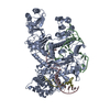
- Structure visualization
| Structure viewer | Molecule: MolmilJmol/JSmol |
|---|
- Downloads & links
-Download
| PDBx/mmCIF format | 1jl5.cif.gz | 94.6 KB | Display | PDBx/mmCIF format |
|---|---|---|---|---|
| PDB format | pdb1jl5.ent.gz | 68.7 KB | Display | PDB format |
| PDBx/mmJSON format | 1jl5.json.gz | Tree view | PDBx/mmJSON format | |
| Others |  Other downloads Other downloads |
-Validation report
| Arichive directory | https://data.pdbj.org/pub/pdb/validation_reports/jl/1jl5ftp://data.pdbj.org/pub/pdb/validation_reports/jl/1jl5 | HTTPS FTP |
|---|
-Related structure data
| Related structure data |  1g9uSC S: Starting model for refinement C: citing same article ( |
|---|---|
| Similar structure data |


-Links
 PDBj
PDBj


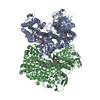
- Assembly
| Deposited unit | 
| |||||||||
|---|---|---|---|---|---|---|---|---|---|---|
| 1 | 
| |||||||||
| Unit cell |
| |||||||||
| Components on special symmetry positions |
| |||||||||
| Details | potentially a tetramer, same as in 1G9U |
-Components
| #1: Protein | Mass: 51588.652 Da / Num. of mol.: 1 Source method: isolated from a genetically manipulated source Source: (gene. exp.) Yersinia pestis (bacteria) / Gene: YopM / Species (production host): Escherichia coli / Production host: Escherichia coli BL21(DE3) (bacteria) / Strain (production host): BL21DE3 / References: UniProt: P17778 | ||
|---|---|---|---|
| #2: Chemical | ChemComp-CA /   Mass: 40.078 Da / Num. of mol.: 5 / Source method: obtained synthetically / Formula: Ca Mass: 40.078 Da / Num. of mol.: 5 / Source method: obtained synthetically / Formula: Ca#3: Water | ChemComp-HOH / | Water Mass: 18.015 Da / Num. of mol.: 339 / Source method: isolated from a natural source / Formula: H2O Mass: 18.015 Da / Num. of mol.: 339 / Source method: isolated from a natural source / Formula: H2O |
-Experimental details
-Experiment
| Experiment | Method: X-RAY DIFFRACTION / Number of used crystals: 1 |
|---|
- Sample preparation
| Crystal grow | Temperature: 300 K / Method: vapor diffusion, hanging drop / pH: 6 Details: 12% isopropanol, 20% ethylene glycol, 0.1M MES, 0.25 M Ca(OAc)2, pH 6.0, VAPOR DIFFUSION, HANGING DROP, temperature 300K | ||||||||||||||||||||||||||||||||||||
|---|---|---|---|---|---|---|---|---|---|---|---|---|---|---|---|---|---|---|---|---|---|---|---|---|---|---|---|---|---|---|---|---|---|---|---|---|---|
| Crystal grow | *PLUS Method: vapor diffusionDetails: Evdokimov, A.G., (2000) Acta Crystallog., D56, 1676. | ||||||||||||||||||||||||||||||||||||
| Components of the solutions | *PLUS
|
-Data collection
| Diffraction | Mean temperature: 100 K |
|---|---|
| Diffraction source | Source: SYNCHROTRON / Site: NSLS  / Beamline: X9B / Wavelength: 0.971 Å / Beamline: X9B / Wavelength: 0.971 Å |
| Detector | Type: ADSC QUANTUM 4 / Detector: CCD / Date: Jan 1, 2001 / Details: multilayer mirrors |
| Radiation | Monochromator: Si crystal (X9B) / Protocol: SINGLE WAVELENGTH / Monochromatic (M) / Laue (L): M / Scattering type: x-ray |
| Radiation wavelength | Wavelength: 0.971 Å / Relative weight: 1 |
| Reflection | Resolution: 2.1→50 Å / Num. all: 58456 / Num. obs: 58456 / % possible obs: 97.3 % / Observed criterion σ(F): 0 / Observed criterion σ(I): 0 / Redundancy: 4.9 % / Biso Wilson estimate: 18 Å2 / Rmerge(I) obs: 0.04 / Net I/σ(I): 25.2 |
| Reflection shell | Resolution: 2.1→2.15 Å / Redundancy: 3.3 % / Rmerge(I) obs: 0.23 / Mean I/σ(I) obs: 4.2 / Num. unique all: 3666 / % possible all: 90.2 |
| Reflection | *PLUS Lowest resolution: 50 Å |
| Reflection shell | *PLUS % possible obs: 92.1 % / Mean I/σ(I) obs: 3.5 |
- Processing
| Software |
| ||||||||||||||||||||||||||||||||||||||||
|---|---|---|---|---|---|---|---|---|---|---|---|---|---|---|---|---|---|---|---|---|---|---|---|---|---|---|---|---|---|---|---|---|---|---|---|---|---|---|---|---|---|
| Refinement | Method to determine structure: MOLECULAR REPLACEMENT Starting model: 1G9U Resolution: 2.1→70.89 Å / Rfactor Rfree error: 0.004 / Data cutoff high absF: 137581.37 / Data cutoff low absF: 0 / Isotropic thermal model: RESTRAINED / Cross valid method: THROUGHOUT / σ(F): 0 / Stereochemistry target values: Engh & Huber
| ||||||||||||||||||||||||||||||||||||||||
| Solvent computation | Solvent model: FLAT MODEL / Bsol: 43.8124 Å2 / ksol: 0.394897 e/Å3 | ||||||||||||||||||||||||||||||||||||||||
| Displacement parameters | Biso mean: 32.3 Å2
| ||||||||||||||||||||||||||||||||||||||||
| Refine analyze |
| ||||||||||||||||||||||||||||||||||||||||
| Refinement step | Cycle: LAST / Resolution: 2.1→70.89 Å
| ||||||||||||||||||||||||||||||||||||||||
| Refine LS restraints |
| ||||||||||||||||||||||||||||||||||||||||
| LS refinement shell | Resolution: 2.1→2.23 Å / Rfactor Rfree error: 0.014 / Total num. of bins used: 6
| ||||||||||||||||||||||||||||||||||||||||
| Xplor file |
| ||||||||||||||||||||||||||||||||||||||||
| Software | *PLUS Name: CNS / Version: 0.9 / Classification: refinement | ||||||||||||||||||||||||||||||||||||||||
| Refinement | *PLUS σ(F): 0 / % reflection Rfree: 5 % / Rfactor obs: 0.22 / Rfactor Rfree: 0.24 / Rfactor Rwork: 0.22 | ||||||||||||||||||||||||||||||||||||||||
| Solvent computation | *PLUS | ||||||||||||||||||||||||||||||||||||||||
| Displacement parameters | *PLUS Biso mean: 32.3 Å2 | ||||||||||||||||||||||||||||||||||||||||
| Refine LS restraints | *PLUS
| ||||||||||||||||||||||||||||||||||||||||
| LS refinement shell | *PLUS Rfactor Rfree: 0.285 / % reflection Rfree: 4.9 % / Rfactor Rwork: 0.27 |
