 Movie
Movie Controller

[English] 日本語
 Yorodumi
- PDB-1g9u: CRYSTAL STRUCTURE OF YOPM-LEUCINE RICH EFFECTOR PROTEIN FROM YERS... -
+ Open data

- Basic information
| Entry | Database: PDB / ID: 1g9u | ||||||
|---|---|---|---|---|---|---|---|
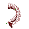
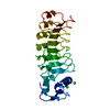
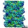
| Title | CRYSTAL STRUCTURE OF YOPM-LEUCINE RICH EFFECTOR PROTEIN FROM YERSINIA PESTIS | ||||||
 Components Components | OUTER PROTEIN YOPM | ||||||
 Keywords Keywords |  TOXIN TOXIN | ||||||
| Function / homology |  Function and homology information Function and homology information | ||||||
| Biological species |   Yersinia pestis (bacteria) Yersinia pestis (bacteria) | ||||||
| Method | X-RAY DIFFRACTION / SYNCHROTRON / MIR / Resolution: 2.35 Å | ||||||
 Authors Authors | Evdokimov, A.G. / Anderson, D.E. / Routzahn, K.M. / Waugh, D.S. | ||||||
 Citation Citation | Journal: J.Mol.Biol. / Year: 2001 Title: Unusual molecular architecture of the Yersinia pestis cytotoxin YopM: a leucine-rich repeat protein with the shortest repeating unit. Authors: Evdokimov, A.G. / Anderson, D.E. / Routzahn, K.M. / Waugh, D.S. | ||||||
| History |
| ||||||
| Remark 300 | BIOMOLECULE: 1 THIS ENTRY CONTAINS THE CRYSTALLOGRAPHIC ASYMMETRIC UNIT WHICH CONSISTS OF 1 CHAIN(S) ...BIOMOLECULE: 1 THIS ENTRY CONTAINS THE CRYSTALLOGRAPHIC ASYMMETRIC UNIT WHICH CONSISTS OF 1 CHAIN(S). SEE REMARK 350 FOR INFORMATION ON GENERATING THE BIOLOGICAL MOLECULE representing a possible biologically significant oligomerization state. |
- Structure visualization
| Structure viewer | Molecule: MolmilJmol/JSmol |
|---|
- Downloads & links
-Download
| PDBx/mmCIF format | 1g9u.cif.gz | 94.1 KB | Display | PDBx/mmCIF format |
|---|---|---|---|---|
| PDB format | pdb1g9u.ent.gz | 68.9 KB | Display | PDB format |
| PDBx/mmJSON format | 1g9u.json.gz | Tree view | PDBx/mmJSON format | |
| Others |  Other downloads Other downloads |
-Validation report
| Arichive directory | https://data.pdbj.org/pub/pdb/validation_reports/g9/1g9uftp://data.pdbj.org/pub/pdb/validation_reports/g9/1g9u | HTTPS FTP |
|---|
-Related structure data


-Links
 PDBj
PDBj


- Assembly
| Deposited unit | 
| ||||||||||||||||||
|---|---|---|---|---|---|---|---|---|---|---|---|---|---|---|---|---|---|---|---|
| 1 | 
| ||||||||||||||||||
| Unit cell |
| ||||||||||||||||||
| Components on special symmetry positions |
| ||||||||||||||||||
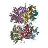
| Details | Putative biological assembly is a tetramer. |
-Components
| #1: Protein | Mass: 51588.652 Da / Num. of mol.: 1 Source method: isolated from a genetically manipulated source Source: (gene. exp.) Yersinia pestis (bacteria) / Gene: YOPM / Species (production host): Escherichia coli / Production host: Escherichia coli BL21(DE3) (bacteria) / Strain (production host): BL21DE3 / References: UniProt: P17778 | ||||||
|---|---|---|---|---|---|---|---|
| #2: Chemical | ChemComp-ACT / Acetate  Mass: 59.044 Da / Num. of mol.: 9 / Source method: obtained synthetically / Formula: C2H3O2 Mass: 59.044 Da / Num. of mol.: 9 / Source method: obtained synthetically / Formula: C2H3O2#3: Chemical |   Mass: 40.078 Da / Num. of mol.: 2 / Source method: obtained synthetically / Formula: Ca Mass: 40.078 Da / Num. of mol.: 2 / Source method: obtained synthetically / Formula: Ca#4: Chemical | ChemComp-HG / Mercury (element)  Mass: 200.590 Da / Num. of mol.: 8 / Source method: obtained synthetically / Formula: Hg Mass: 200.590 Da / Num. of mol.: 8 / Source method: obtained synthetically / Formula: Hg#5: Water | ChemComp-HOH / | Water Mass: 18.015 Da / Num. of mol.: 236 / Source method: isolated from a natural source / Formula: H2O Mass: 18.015 Da / Num. of mol.: 236 / Source method: isolated from a natural source / Formula: H2O |
-Experimental details
-Experiment
| Experiment | Method: X-RAY DIFFRACTION / Number of used crystals: 1 |
|---|
- Sample preparation
| Crystal | Density Matthews: 2.94 Å3/Da / Density % sol: 58.14 % | ||||||||||||||||||||||||||||||||||||
|---|---|---|---|---|---|---|---|---|---|---|---|---|---|---|---|---|---|---|---|---|---|---|---|---|---|---|---|---|---|---|---|---|---|---|---|---|---|
| Crystal grow | Method: vapor diffusion, hanging drop / pH: 6 Details: 10% isopropanol 0.1 M MES, 0.2-0.4 M Ca(OAc)2, pH 6.0, VAPOR DIFFUSION, HANGING DROP, temperature 321.0K | ||||||||||||||||||||||||||||||||||||
| Crystal grow | *PLUS Method: vapor diffusionDetails: Evdokimov, A.G., (2000) Acta Crystallog., D56, 1676. | ||||||||||||||||||||||||||||||||||||
| Components of the solutions | *PLUS
|
-Data collection
| Diffraction | Mean temperature: 100 K |
|---|---|
| Diffraction source | Source: SYNCHROTRON / Site: NSLS  / Beamline: X9B / Wavelength: 0.9879 Å / Beamline: X9B / Wavelength: 0.9879 Å |
| Detector | Type: ADSC QUANTUM 4 / Detector: CCD / Date: Mar 22, 2000 / Details: synchrotron Si monochromator + Osmic mirrors |
| Radiation | Monochromator: Si 111 / Protocol: SINGLE WAVELENGTH / Monochromatic (M) / Laue (L): M / Scattering type: x-ray |
| Radiation wavelength | Wavelength: 0.9879 Å / Relative weight: 1 |
| Reflection | Resolution: 2.32→74 Å / Num. all: 49157 / Num. obs: 39896 / % possible obs: 81 % / Observed criterion σ(F): 4 / Observed criterion σ(I): 4 / Redundancy: 1.9 % / Biso Wilson estimate: 18 Å2 / Rmerge(I) obs: 0.1 / Net I/σ(I): 9 |
| Reflection shell | Resolution: 2.32→2.45 Å / Redundancy: 1.8 % / Rmerge(I) obs: 0.43 / Mean I/σ(I) obs: 2.2 / Num. unique all: 7609 / % possible all: 96.4 |
| Reflection | *PLUS Lowest resolution: 74 Å |
- Processing
| Software |
| |||||||||||||||||||||||||
|---|---|---|---|---|---|---|---|---|---|---|---|---|---|---|---|---|---|---|---|---|---|---|---|---|---|---|
| Refinement | Method to determine structure: MIR Starting model: new structure Resolution: 2.35→100 Å / Isotropic thermal model: isotropic per atom / Cross valid method: random 5% throughout the refinement / σ(F): 0 / σ(I): 0 / Stereochemistry target values: Engh & Huber
| |||||||||||||||||||||||||
| Displacement parameters | Biso mean: 33 Å2 | |||||||||||||||||||||||||
| Refinement step | Cycle: LAST / Resolution: 2.35→100 Å
| |||||||||||||||||||||||||
| Refine LS restraints |
| |||||||||||||||||||||||||
| LS refinement shell | Resolution: 2.35→100 Å / Rfactor Rfree error: 0.02
| |||||||||||||||||||||||||
| Software | *PLUS Name: SHELXL-97 / Classification: refinement | |||||||||||||||||||||||||
| Refinement | *PLUS Lowest resolution: 100 Å / σ(F): 0 / % reflection Rfree: 5 % / Rfactor all: 0.21 / Rfactor obs: 0.17 / Rfactor Rfree: 0.23 / Rfactor Rwork: 0.17 | |||||||||||||||||||||||||
| Solvent computation | *PLUS | |||||||||||||||||||||||||
| Displacement parameters | *PLUS | |||||||||||||||||||||||||
| Refine LS restraints | *PLUS
|
