 Movie
Movie Controller


+ Open data

- Basic information
| Entry | Database: PDB / ID: 4c4k | ||||||
|---|---|---|---|---|---|---|---|
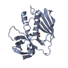
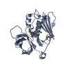
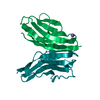
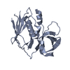
| Title | Crystal structure of the titin M10-Obscurin Ig domain 1 complex | ||||||
 Components Components |
| ||||||
 Keywords Keywords |  TRANSFERASE / SARCOMERE / IMMUNOGLOBULIN DOMAIN / LIMB-GIRDLE MUSCULAR DYSTROPHY TRANSFERASE / SARCOMERE / IMMUNOGLOBULIN DOMAIN / LIMB-GIRDLE MUSCULAR DYSTROPHY | ||||||
| Function / homology |  Function and homology information Function and homology informationprotein localization to M-band / sarcomerogenesis / structural molecule activity conferring elasticity / telethonin binding / skeletal muscle myosin thick filament assembly / phosphatidylinositol-5-phosphate binding / cardiac myofibril assembly / muscle alpha-actinin binding / detection of muscle stretch / cardiac muscle tissue morphogenesis ...protein localization to M-band / sarcomerogenesis / structural molecule activity conferring elasticity / telethonin binding / skeletal muscle myosin thick filament assembly / phosphatidylinositol-5-phosphate binding / cardiac myofibril assembly / muscle alpha-actinin binding / detection of muscle stretch / cardiac muscle tissue morphogenesis / regulation of catalytic activity / phosphatidylinositol-3-phosphate binding / phosphatidylinositol-3,4-bisphosphate binding / cardiac muscle hypertrophy / mitotic chromosome condensation / Striated Muscle Contraction / actinin binding / M band / I band / phosphatidylinositol-4-phosphate binding / cardiac muscle cell development / regulation of protein kinase activity / regulation of small GTPase mediated signal transduction / structural constituent of muscle / ankyrin binding / sarcomere organization / myofibril / NRAGE signals death through JNK / RHOQ GTPase cycle / phosphatidylinositol-3,4,5-trisphosphate binding / skeletal muscle thin filament assembly / striated muscle thin filament / RHOA GTPase cycle / striated muscle contraction / cardiac muscle contraction / titin binding / protein kinase A signaling / phosphatidylinositol-4,5-bisphosphate binding / guanyl-nucleotide exchange factor activity / condensed nuclear chromosome / muscle contraction / positive regulation of protein secretion / sarcolemma / Z disc / response to calcium ion / : / G alpha (12/13) signalling events / actin filament binding / Platelet degranulation / protein tyrosine kinase activity / protease binding / nuclear body / calmodulin binding / non-specific serine/threonine protein kinase / phosphorylation / protein serine kinase activity / protein serine/threonine kinase activity / calcium ion binding / positive regulation of gene expression / protein kinase binding / enzyme binding / extracellular exosome / extracellular region / ATP binding / identical protein binding / metal ion binding / plasma membrane / cytosolSimilarity search - Function | ||||||
| Biological species |  HOMO SAPIENS (human) HOMO SAPIENS (human) | ||||||
| Method | X-RAY DIFFRACTION / SYNCHROTRON / MOLECULAR REPLACEMENT / Resolution: 1.95 Å | ||||||
 Authors Authors | Pernigo, S. / Fukuzawa, A. / Gautel, M. / Steiner, R.A. | ||||||
 Citation Citation | Journal: J.Mol.Biol. / Year: 2015 Title: The Crystal Structure of the Human Titin:Obscurin Complex Reveals a Conserved Yet Specific Muscle M-Band Zipper Module. Authors: Pernigo, S. / Fukuzawa, A. / Pandini, A. / Holt, M. / Kleinjung, J. / Gautel, M. / Steiner, R.A. | ||||||
| History |
|
- Structure visualization
| Structure viewer | Molecule: MolmilJmol/JSmol |
|---|
- Downloads & links
-Download
| PDBx/mmCIF format | 4c4k.cif.gz | 93.9 KB | Display | PDBx/mmCIF format |
|---|---|---|---|---|
| PDB format | pdb4c4k.ent.gz | 72 KB | Display | PDB format |
| PDBx/mmJSON format | 4c4k.json.gz | Tree view | PDBx/mmJSON format | |
| Others |  Other downloads Other downloads |
-Validation report
| Arichive directory | https://data.pdbj.org/pub/pdb/validation_reports/c4/4c4kftp://data.pdbj.org/pub/pdb/validation_reports/c4/4c4k | HTTPS FTP |
|---|
-Related structure data
| Related structure data |  4uowC  2wp3S S: Starting model for refinement C: citing same article ( |
|---|---|
| Similar structure data |


-Links
 PDBj
PDBj


- Assembly
| Deposited unit | 
| ||||||||
|---|---|---|---|---|---|---|---|---|---|
| 1 |
| ||||||||
| Unit cell |
|
-Components
| #1: Protein | / OBSCURIN-RHOGEF / OBSCURIN-MYOSIN LIGHT CHAIN KINASE / OBSCURIN-MLCK Mass: 10373.669 Da / Num. of mol.: 1 / Fragment: FIRST IG DOMAIN, RESIDUES 9-103 Source method: isolated from a genetically manipulated source Source: (gene. exp.) HOMO SAPIENS (human) / Production host:  ESCHERICHIA COLI (E. coli) / Strain (production host): BL21(DE3) / Variant (production host): ROSETTA 2 ESCHERICHIA COLI (E. coli) / Strain (production host): BL21(DE3) / Variant (production host): ROSETTA 2References: UniProt: Q5VST9, non-specific serine/threonine protein kinase | ||||
|---|---|---|---|---|---|
| #2: Protein | / CONNECTIN / RHABDOMYOSARCOMA ANTIGEN MU-RMS-40.14 Mass: 10981.231 Da / Num. of mol.: 1 / Fragment: M10 DOMAIN, RESIDUES 34252-34350 Source method: isolated from a genetically manipulated source Source: (gene. exp.) HOMO SAPIENS (human) / Production host: ESCHERICHIA COLI (E. coli) / Strain (production host): BL21(DE3) / Variant (production host): ROSETTA 2References: UniProt: Q8WZ42, non-specific serine/threonine protein kinase | ||||
| #3: Chemical | ChemComp-EDO / Ethylene glycol  Mass: 62.068 Da / Num. of mol.: 12 / Source method: obtained synthetically / Formula: C2H6O2 Mass: 62.068 Da / Num. of mol.: 12 / Source method: obtained synthetically / Formula: C2H6O2#4: Water | ChemComp-HOH / | Water Mass: 18.015 Da / Num. of mol.: 103 / Source method: isolated from a natural source / Formula: H2O Mass: 18.015 Da / Num. of mol.: 103 / Source method: isolated from a natural source / Formula: H2OSequence details | INITIAL GSS IS DERIVED FROM VECTOR AFTER TEV CLEAVAGE | |
-Experimental details
-Experiment
| Experiment | Method: X-RAY DIFFRACTION / Number of used crystals: 1 |
|---|
- Sample preparation
| Crystal | Density Matthews: 2.34 Å3/Da / Density % sol: 47.5 % / Description: NONE |
|---|---|
| Crystal grow | pH: 8.5 Details: 0.1 M TRIS-HCL PH 8.5, 0.2 M SODIUM ACETATE, 30% PEG4000 |
-Data collection
| Diffraction | Mean temperature: 100 K |
|---|---|
| Diffraction source | Source: SYNCHROTRON / Site: Diamond  / Beamline: I24 / Wavelength: 0.9686 / Beamline: I24 / Wavelength: 0.9686 |
| Detector | Type: DECTRIS PILATUS 6M / Detector: PIXEL / Date: Dec 6, 2012 / Details: MIRRORS |
| Radiation | Monochromator: SI(111) / Protocol: SINGLE WAVELENGTH / Monochromatic (M) / Laue (L): M / Scattering type: x-ray |
| Radiation wavelength | Wavelength: 0.9686 Å / Relative weight: 1 |
| Reflection | Resolution: 1.95→35.7 Å / Num. obs: 15005 / % possible obs: 99 % / Observed criterion σ(I): 0 / Redundancy: 3.3 % / Biso Wilson estimate: 31.76 Å2 / Rmerge(I) obs: 0.05 / Net I/σ(I): 12.8 |
| Reflection shell | Resolution: 1.95→2 Å / Redundancy: 3.1 % / Rmerge(I) obs: 0.62 / Mean I/σ(I) obs: 2 / % possible all: 98.5 |
- Processing
| Software |
| |||||||||||||||||||||||||||||||||||||||||||||||||||||||||||||||||||||||||||||||||||||||||||||||||||||||||||||||||||||||||||||||||||||||||||||||||||||||||||||||||||||||||||||||||||||||||||||||||||||||||||||||||||||||||||||||||||||||||||||||||||||||||||||||||||||||||||||||||||||||||||||||||||||||||||||||||||||||||||||||||||||||||||||||||||||||||||||||||||||||||||||||||||||||||||||||||||||||||||||||||||||||||||||||||||||||||||||||||||||||||||||||||||||||||||||||||||||||||||||||||||||||||||||||||||||||||||||||||||||||||||||
|---|---|---|---|---|---|---|---|---|---|---|---|---|---|---|---|---|---|---|---|---|---|---|---|---|---|---|---|---|---|---|---|---|---|---|---|---|---|---|---|---|---|---|---|---|---|---|---|---|---|---|---|---|---|---|---|---|---|---|---|---|---|---|---|---|---|---|---|---|---|---|---|---|---|---|---|---|---|---|---|---|---|---|---|---|---|---|---|---|---|---|---|---|---|---|---|---|---|---|---|---|---|---|---|---|---|---|---|---|---|---|---|---|---|---|---|---|---|---|---|---|---|---|---|---|---|---|---|---|---|---|---|---|---|---|---|---|---|---|---|---|---|---|---|---|---|---|---|---|---|---|---|---|---|---|---|---|---|---|---|---|---|---|---|---|---|---|---|---|---|---|---|---|---|---|---|---|---|---|---|---|---|---|---|---|---|---|---|---|---|---|---|---|---|---|---|---|---|---|---|---|---|---|---|---|---|---|---|---|---|---|---|---|---|---|---|---|---|---|---|---|---|---|---|---|---|---|---|---|---|---|---|---|---|---|---|---|---|---|---|---|---|---|---|---|---|---|---|---|---|---|---|---|---|---|---|---|---|---|---|---|---|---|---|---|---|---|---|---|---|---|---|---|---|---|---|---|---|---|---|---|---|---|---|---|---|---|---|---|---|---|---|---|---|---|---|---|---|---|---|---|---|---|---|---|---|---|---|---|---|---|---|---|---|---|---|---|---|---|---|---|---|---|---|---|---|---|---|---|---|---|---|---|---|---|---|---|---|---|---|---|---|---|---|---|---|---|---|---|---|---|---|---|---|---|---|---|---|---|---|---|---|---|---|---|---|---|---|---|---|---|---|---|---|---|---|---|---|---|---|---|---|---|---|---|---|---|---|---|---|---|---|---|---|---|---|---|---|---|---|---|---|---|---|---|---|---|---|---|---|---|---|---|---|---|---|---|---|---|---|---|---|---|---|---|---|---|---|---|---|---|---|---|---|---|---|---|---|---|---|---|---|---|---|---|---|---|---|---|---|---|---|---|---|---|---|---|---|---|---|---|---|---|---|---|---|---|---|---|---|---|---|---|---|---|---|---|---|---|---|---|---|---|---|---|---|---|---|---|---|---|---|---|---|---|---|---|---|---|---|---|---|---|---|---|---|---|---|---|---|---|---|---|---|---|---|---|---|---|---|---|---|---|---|---|---|---|
| Refinement | Method to determine structure: MOLECULAR REPLACEMENT Starting model: PDB ENTRY 2WP3 Resolution: 1.95→17.48 Å / Cor.coef. Fo:Fc: 0.9549 / Cor.coef. Fo:Fc free: 0.9429 / SU R Cruickshank DPI: 0.146 / Cross valid method: THROUGHOUT / σ(F): 0 / SU R Blow DPI: 0.155 / SU Rfree Blow DPI: 0.138 / SU Rfree Cruickshank DPI: 0.135
| |||||||||||||||||||||||||||||||||||||||||||||||||||||||||||||||||||||||||||||||||||||||||||||||||||||||||||||||||||||||||||||||||||||||||||||||||||||||||||||||||||||||||||||||||||||||||||||||||||||||||||||||||||||||||||||||||||||||||||||||||||||||||||||||||||||||||||||||||||||||||||||||||||||||||||||||||||||||||||||||||||||||||||||||||||||||||||||||||||||||||||||||||||||||||||||||||||||||||||||||||||||||||||||||||||||||||||||||||||||||||||||||||||||||||||||||||||||||||||||||||||||||||||||||||||||||||||||||||||||||||||||
| Displacement parameters |
| |||||||||||||||||||||||||||||||||||||||||||||||||||||||||||||||||||||||||||||||||||||||||||||||||||||||||||||||||||||||||||||||||||||||||||||||||||||||||||||||||||||||||||||||||||||||||||||||||||||||||||||||||||||||||||||||||||||||||||||||||||||||||||||||||||||||||||||||||||||||||||||||||||||||||||||||||||||||||||||||||||||||||||||||||||||||||||||||||||||||||||||||||||||||||||||||||||||||||||||||||||||||||||||||||||||||||||||||||||||||||||||||||||||||||||||||||||||||||||||||||||||||||||||||||||||||||||||||||||||||||||||
| Refine analyze | Luzzati coordinate error obs: 0.222 Å | |||||||||||||||||||||||||||||||||||||||||||||||||||||||||||||||||||||||||||||||||||||||||||||||||||||||||||||||||||||||||||||||||||||||||||||||||||||||||||||||||||||||||||||||||||||||||||||||||||||||||||||||||||||||||||||||||||||||||||||||||||||||||||||||||||||||||||||||||||||||||||||||||||||||||||||||||||||||||||||||||||||||||||||||||||||||||||||||||||||||||||||||||||||||||||||||||||||||||||||||||||||||||||||||||||||||||||||||||||||||||||||||||||||||||||||||||||||||||||||||||||||||||||||||||||||||||||||||||||||||||||||
| Refinement step | Cycle: LAST / Resolution: 1.95→17.48 Å
| |||||||||||||||||||||||||||||||||||||||||||||||||||||||||||||||||||||||||||||||||||||||||||||||||||||||||||||||||||||||||||||||||||||||||||||||||||||||||||||||||||||||||||||||||||||||||||||||||||||||||||||||||||||||||||||||||||||||||||||||||||||||||||||||||||||||||||||||||||||||||||||||||||||||||||||||||||||||||||||||||||||||||||||||||||||||||||||||||||||||||||||||||||||||||||||||||||||||||||||||||||||||||||||||||||||||||||||||||||||||||||||||||||||||||||||||||||||||||||||||||||||||||||||||||||||||||||||||||||||||||||||
| Refine LS restraints |
| |||||||||||||||||||||||||||||||||||||||||||||||||||||||||||||||||||||||||||||||||||||||||||||||||||||||||||||||||||||||||||||||||||||||||||||||||||||||||||||||||||||||||||||||||||||||||||||||||||||||||||||||||||||||||||||||||||||||||||||||||||||||||||||||||||||||||||||||||||||||||||||||||||||||||||||||||||||||||||||||||||||||||||||||||||||||||||||||||||||||||||||||||||||||||||||||||||||||||||||||||||||||||||||||||||||||||||||||||||||||||||||||||||||||||||||||||||||||||||||||||||||||||||||||||||||||||||||||||||||||||||||
| LS refinement shell | Resolution: 1.95→2.08 Å / Total num. of bins used: 8
| |||||||||||||||||||||||||||||||||||||||||||||||||||||||||||||||||||||||||||||||||||||||||||||||||||||||||||||||||||||||||||||||||||||||||||||||||||||||||||||||||||||||||||||||||||||||||||||||||||||||||||||||||||||||||||||||||||||||||||||||||||||||||||||||||||||||||||||||||||||||||||||||||||||||||||||||||||||||||||||||||||||||||||||||||||||||||||||||||||||||||||||||||||||||||||||||||||||||||||||||||||||||||||||||||||||||||||||||||||||||||||||||||||||||||||||||||||||||||||||||||||||||||||||||||||||||||||||||||||||||||||||
| Refinement TLS params. | Method: refined / Refine-ID: X-RAY DIFFRACTION
| |||||||||||||||||||||||||||||||||||||||||||||||||||||||||||||||||||||||||||||||||||||||||||||||||||||||||||||||||||||||||||||||||||||||||||||||||||||||||||||||||||||||||||||||||||||||||||||||||||||||||||||||||||||||||||||||||||||||||||||||||||||||||||||||||||||||||||||||||||||||||||||||||||||||||||||||||||||||||||||||||||||||||||||||||||||||||||||||||||||||||||||||||||||||||||||||||||||||||||||||||||||||||||||||||||||||||||||||||||||||||||||||||||||||||||||||||||||||||||||||||||||||||||||||||||||||||||||||||||||||||||||
| Refinement TLS group |
|
