 Movie
Movie Controller


+ Open data

- Basic information
| Entry | Database: PDB / ID: 4bwk | ||||||
|---|---|---|---|---|---|---|---|
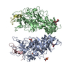
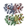
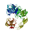
| Title | Structure of Neurospora crassa PAN3 pseudokinase | ||||||
 Components Components | PAB-DEPENDENT POLY(A)-SPECIFIC RIBONUCLEASE SUBUNIT PAN-3 | ||||||
 Keywords Keywords |  GENE REGULATION / DEADENYLASE / PAN2 / MIRNA / MRNA DECAY GENE REGULATION / DEADENYLASE / PAN2 / MIRNA / MRNA DECAY | ||||||
| Function / homology |  Function and homology information Function and homology informationPAN complex / nuclear-transcribed mRNA poly(A) tail shortening / poly(A) binding / P-body / mRNA processing / ATP binding / metal ion bindingSimilarity search - Function | ||||||
| Biological species |  NEUROSPORA CRASSA (fungus) NEUROSPORA CRASSA (fungus) | ||||||
| Method | X-RAY DIFFRACTION / SYNCHROTRON / MOLECULAR REPLACEMENT / Resolution: 3.3 Å | ||||||
 Authors Authors | Christie, M. / Boland, A. / Huntzinger, E. / Weichenrieder, O. / Izaurralde, E. | ||||||
 Citation Citation | Journal: Mol.Cell / Year: 2013 Title: Structure of the Pan3 Pseudokinase Reveals the Basis for Interactions with the Pan2 Deadenylase and the Gw182 Proteins Authors: Christie, M. / Boland, A. / Huntzinger, E. / Weichenrieder, O. / Izaurralde, E. | ||||||
| History |
| ||||||
| Remark 650 | HELIX DETERMINATION METHOD: AUTHOR PROVIDED. | ||||||
| Remark 700 | SHEET DETERMINATION METHOD: AUTHOR PROVIDED. |
- Structure visualization
| Structure viewer | Molecule: MolmilJmol/JSmol |
|---|
- Downloads & links
-Download
| PDBx/mmCIF format | 4bwk.cif.gz | 331.9 KB | Display | PDBx/mmCIF format |
|---|---|---|---|---|
| PDB format | pdb4bwk.ent.gz | 277.2 KB | Display | PDB format |
| PDBx/mmJSON format | 4bwk.json.gz | Tree view | PDBx/mmJSON format | |
| Others |  Other downloads Other downloads |
-Validation report
| Arichive directory | https://data.pdbj.org/pub/pdb/validation_reports/bw/4bwkftp://data.pdbj.org/pub/pdb/validation_reports/bw/4bwk | HTTPS FTP |
|---|
-Related structure data


-Links
 PDBj
PDBj

- Assembly
| Deposited unit | 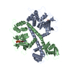
| ||||||||
|---|---|---|---|---|---|---|---|---|---|
| 1 |
| ||||||||
| Unit cell |
| ||||||||
| Noncrystallographic symmetry (NCS) | NCS oper: (Code: given Matrix: (-0.998, 0.014, 0.057), Vector : |
-Components
| #1: Protein | Mass: 49269.078 Da / Num. of mol.: 2 Fragment: PSEUDOKINASE DOMAIN, COILED COIL, CTERMINAL KNOB, RESIDUES 234-656 Source method: isolated from a genetically manipulated source Source: (gene. exp.) NEUROSPORA CRASSA (fungus) / Plasmid: PNEA-NPM / Production host:  ESCHERICHIA COLI (E. coli) / Strain (production host): BL21(DE3) / References: UniProt: Q7SDP4 ESCHERICHIA COLI (E. coli) / Strain (production host): BL21(DE3) / References: UniProt: Q7SDP4#2: Chemical |   Mass: 523.247 Da / Num. of mol.: 2 / Source method: obtained synthetically / Formula: C10H16N5O12P3S / Comment: ATP-gamma-S, energy-carrying molecule analogue*YM Mass: 523.247 Da / Num. of mol.: 2 / Source method: obtained synthetically / Formula: C10H16N5O12P3S / Comment: ATP-gamma-S, energy-carrying molecule analogue*YM |
|---|
-Experimental details
-Experiment
| Experiment | Method: X-RAY DIFFRACTION / Number of used crystals: 1 |
|---|
- Sample preparation
| Crystal | Density Matthews: 2.65 Å3/Da / Density % sol: 53.5 % / Description: NONE |
|---|---|
| Crystal grow | pH: 7.5 / Details: 0.1 M HEPES PH 7.5, 7% PEG 6000, 6% ISOPROPANOL |
-Data collection
| Diffraction | Mean temperature: 100 K |
|---|---|
| Diffraction source | Source: SYNCHROTRON / Site: SLS  / Beamline: X10SA / Wavelength: 1.00001 / Beamline: X10SA / Wavelength: 1.00001 |
| Detector | Type: DECTRIS PILATUS 6M / Detector: PIXEL / Date: Mar 26, 2012 / Details: MIRRORS |
| Radiation | Monochromator: SI(111) / Protocol: SINGLE WAVELENGTH / Monochromatic (M) / Laue (L): M / Scattering type: x-ray |
| Radiation wavelength | Wavelength: 1.00001 Å / Relative weight: 1 |
| Reflection | Resolution: 3.3→46.03 Å / Num. obs: 15611 / % possible obs: 99.9 % / Observed criterion σ(I): -3 / Redundancy: 7.9 % / Biso Wilson estimate: 117.45 Å2 / Rmerge(I) obs: 0.06 / Net I/σ(I): 19.9 |
| Reflection shell | Resolution: 3.3→3.48 Å / Redundancy: 7.7 % / Rmerge(I) obs: 0.77 / Mean I/σ(I) obs: 3 / % possible all: 99.8 |
- Processing
| Software |
| |||||||||||||||||||||||||||||||||||||||||||||||||||||||||||||||||||||||||||
|---|---|---|---|---|---|---|---|---|---|---|---|---|---|---|---|---|---|---|---|---|---|---|---|---|---|---|---|---|---|---|---|---|---|---|---|---|---|---|---|---|---|---|---|---|---|---|---|---|---|---|---|---|---|---|---|---|---|---|---|---|---|---|---|---|---|---|---|---|---|---|---|---|---|---|---|---|
| Refinement | Method to determine structure: MOLECULAR REPLACEMENT / Resolution: 3.3→46.027 Å / SU ML: 0.4 / σ(F): 1.36 / Phase error: 37.74 / Stereochemistry target values: ML
| |||||||||||||||||||||||||||||||||||||||||||||||||||||||||||||||||||||||||||
| Solvent computation | Shrinkage radii: 0.9 Å / VDW probe radii: 1.11 Å / Solvent model: FLAT BULK SOLVENT MODEL | |||||||||||||||||||||||||||||||||||||||||||||||||||||||||||||||||||||||||||
| Refinement step | Cycle: LAST / Resolution: 3.3→46.027 Å
| |||||||||||||||||||||||||||||||||||||||||||||||||||||||||||||||||||||||||||
| Refine LS restraints |
| |||||||||||||||||||||||||||||||||||||||||||||||||||||||||||||||||||||||||||
| LS refinement shell |
| |||||||||||||||||||||||||||||||||||||||||||||||||||||||||||||||||||||||||||
| Refinement TLS params. | Method: refined / Refine-ID: X-RAY DIFFRACTION
| |||||||||||||||||||||||||||||||||||||||||||||||||||||||||||||||||||||||||||
| Refinement TLS group |
|
