 Movie
Movie Controller

[English] 日本語
 Yorodumi
- PDB-4bpt: Structural and thermodynamic insight into phenylalanine hydroxyla... -
+ Open data

- Basic information
| Entry | Database: PDB / ID: 4bpt | ||||||
|---|---|---|---|---|---|---|---|
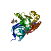
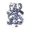
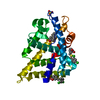
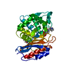
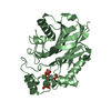
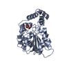
| Title | Structural and thermodynamic insight into phenylalanine hydroxylase from the human pathogen Legionella pneumophila | ||||||
 Components Components | PHENYLALANINE-4-HYDROXYLASE (PAH) (PHE-4-MONOOXYGENASE) | ||||||
 Keywords Keywords |  OXIDOREDUCTASE / PYOMELANIN SYNTHESIS / THERMOSTABILITY / AGGREGATION OXIDOREDUCTASE / PYOMELANIN SYNTHESIS / THERMOSTABILITY / AGGREGATION | ||||||
| Function / homology |  Function and homology informationphenylalanine 4-monooxygenase / phenylalanine 4-monooxygenase activity / L-phenylalanine catabolic process / iron ion binding Function and homology informationphenylalanine 4-monooxygenase / phenylalanine 4-monooxygenase activity / L-phenylalanine catabolic process / iron ion bindingSimilarity search - Function | ||||||
| Biological species |   LEGIONELLA PNEUMOPHILA (bacteria) LEGIONELLA PNEUMOPHILA (bacteria) | ||||||
| Method | X-RAY DIFFRACTION / SYNCHROTRON / MOLECULAR REPLACEMENT / Resolution: 2.5 Å | ||||||
 Authors Authors | Leiros, H.-K.S. / Flydal, M.I. / Martinez, A. | ||||||
 Citation Citation | Journal: FEBS Open Bio / Year: 2013 Title: Structural and Thermodynamic Insight Into Phenylalanine Hydroxylase from the Human Pathogen Legionella Pneumophila. Authors: Leiros, H.S. / Flydal, M.I. / Martinez, A. | ||||||
| History |
|
- Structure visualization
| Structure viewer | Molecule: MolmilJmol/JSmol |
|---|
- Downloads & links
-Download
| PDBx/mmCIF format | 4bpt.cif.gz | 321.3 KB | Display | PDBx/mmCIF format |
|---|---|---|---|---|
| PDB format | pdb4bpt.ent.gz | 259.8 KB | Display | PDB format |
| PDBx/mmJSON format | 4bpt.json.gz | Tree view | PDBx/mmJSON format | |
| Others |  Other downloads Other downloads |
-Validation report
| Arichive directory | https://data.pdbj.org/pub/pdb/validation_reports/bp/4bptftp://data.pdbj.org/pub/pdb/validation_reports/bp/4bpt | HTTPS FTP |
|---|
-Related structure data
| Related structure data |  2v27S S: Starting model for refinement |
|---|---|
| Similar structure data |


-Links
 PDBj
PDBj
- Assembly
| Deposited unit | 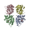
| ||||||||
|---|---|---|---|---|---|---|---|---|---|
| 1 | 
| ||||||||
| 2 | 
| ||||||||
| 3 | 
| ||||||||
| 4 | 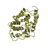
| ||||||||
| Unit cell |
|
-Components
| #1: Protein | Mass: 31418.826 Da / Num. of mol.: 4 Source method: isolated from a genetically manipulated source Source: (gene. exp.) LEGIONELLA PNEUMOPHILA (bacteria) / Production host: ESCHERICHIA COLI (E. coli) / Strain (production host): BL21(DE3) / Variant (production host): CODON PLUS RILReferences: UniProt: I7HM43, UniProt: Q5ZS72*PLUS, phenylalanine 4-monooxygenase#2: Chemical | ChemComp-PEG / Diethylene glycol  Mass: 106.120 Da / Num. of mol.: 6 / Source method: obtained synthetically / Formula: C4H10O3 Mass: 106.120 Da / Num. of mol.: 6 / Source method: obtained synthetically / Formula: C4H10O3#3: Water | ChemComp-HOH / | Water Mass: 18.015 Da / Num. of mol.: 155 / Source method: isolated from a natural source / Formula: H2O Mass: 18.015 Da / Num. of mol.: 155 / Source method: isolated from a natural source / Formula: H2O |
|---|
-Experimental details
-Experiment
| Experiment | Method: X-RAY DIFFRACTION / Number of used crystals: 1 |
|---|
- Sample preparation
| Crystal | Density Matthews: 2.67 Å3/Da / Density % sol: 53.98 % / Description: NONE |
|---|---|
| Crystal grow | Details: 1.6 M NAK2PO4, 4% (V/V) PEG 400 |
-Data collection
| Diffraction | Mean temperature: 100 K |
|---|---|
| Diffraction source | Source: SYNCHROTRON / Site: BESSY  / Beamline: 14.1 / Wavelength: 0.91841 / Beamline: 14.1 / Wavelength: 0.91841 |
| Detector | Type: MARMOSAIC 225 mm CCD / Detector: CCD |
| Radiation | Protocol: SINGLE WAVELENGTH / Monochromatic (M) / Laue (L): M / Scattering type: x-ray |
| Radiation wavelength | Wavelength: 0.91841 Å / Relative weight: 1 |
| Reflection | Resolution: 2.5→25 Å / Num. obs: 41892 / % possible obs: 90.4 % / Observed criterion σ(I): 0 / Redundancy: 2.3 % / Rmerge(I) obs: 0.08 / Net I/σ(I): 0 |
| Reflection shell | Resolution: 2.5→2.64 Å / Redundancy: 2.3 % / Rmerge(I) obs: 0.39 / Mean I/σ(I) obs: 2.2 / % possible all: 94 |
- Processing
| Software |
| ||||||||||||||||||||||||||||||||||||||||||||||||||||||||||||||||||||||||||||||||||||||||||||||||||||||||||||||||||||||||||||||||||||||||||||||||||||||||||||||||||||||||||||||||||||||
|---|---|---|---|---|---|---|---|---|---|---|---|---|---|---|---|---|---|---|---|---|---|---|---|---|---|---|---|---|---|---|---|---|---|---|---|---|---|---|---|---|---|---|---|---|---|---|---|---|---|---|---|---|---|---|---|---|---|---|---|---|---|---|---|---|---|---|---|---|---|---|---|---|---|---|---|---|---|---|---|---|---|---|---|---|---|---|---|---|---|---|---|---|---|---|---|---|---|---|---|---|---|---|---|---|---|---|---|---|---|---|---|---|---|---|---|---|---|---|---|---|---|---|---|---|---|---|---|---|---|---|---|---|---|---|---|---|---|---|---|---|---|---|---|---|---|---|---|---|---|---|---|---|---|---|---|---|---|---|---|---|---|---|---|---|---|---|---|---|---|---|---|---|---|---|---|---|---|---|---|---|---|---|---|
| Refinement | Method to determine structure: MOLECULAR REPLACEMENT Starting model: PDB ENTRY 2V27 Resolution: 2.5→10 Å / Cor.coef. Fo:Fc: 0.89 / Cor.coef. Fo:Fc free: 0.867 / SU B: 12.643 / SU ML: 0.177 / Cross valid method: THROUGHOUT / ESU R: 0.138 / ESU R Free: 0.074 / Stereochemistry target values: MAXIMUM LIKELIHOOD Details: HYDROGENS HAVE BEEN ADDED IN THE RIDING POSITIONS. U VALUES WITH TLS ADDED
| ||||||||||||||||||||||||||||||||||||||||||||||||||||||||||||||||||||||||||||||||||||||||||||||||||||||||||||||||||||||||||||||||||||||||||||||||||||||||||||||||||||||||||||||||||||||
| Solvent computation | Ion probe radii: 0.8 Å / Shrinkage radii: 0.8 Å / VDW probe radii: 1.2 Å / Solvent model: MASK | ||||||||||||||||||||||||||||||||||||||||||||||||||||||||||||||||||||||||||||||||||||||||||||||||||||||||||||||||||||||||||||||||||||||||||||||||||||||||||||||||||||||||||||||||||||||
| Displacement parameters | Biso mean: 37.801 Å2
| ||||||||||||||||||||||||||||||||||||||||||||||||||||||||||||||||||||||||||||||||||||||||||||||||||||||||||||||||||||||||||||||||||||||||||||||||||||||||||||||||||||||||||||||||||||||
| Refinement step | Cycle: LAST / Resolution: 2.5→10 Å
| ||||||||||||||||||||||||||||||||||||||||||||||||||||||||||||||||||||||||||||||||||||||||||||||||||||||||||||||||||||||||||||||||||||||||||||||||||||||||||||||||||||||||||||||||||||||
| Refine LS restraints |
|