 Movie
Movie Controller

[English] 日本語
 Yorodumi
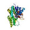
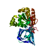
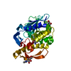
- PDB-3bs4: Crystal structure of uncharacterized protein PH0321 from Pyrococc... -
+ Open data

- Basic information
| Entry | Database: PDB / ID: 3bs4 | ||||||
|---|---|---|---|---|---|---|---|
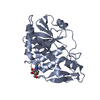
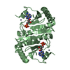
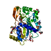
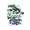
| Title | Crystal structure of uncharacterized protein PH0321 from Pyrococcus horikoshii in complex with an unknown peptide | ||||||
 Components Components |
| ||||||
 Keywords Keywords |  STRUCTURAL GENOMICS / UNKNOWN FUNCTION / PSI-2 / Protein Structure Initiative / New York SGX Research Center for Structural Genomics / NYSGXRC STRUCTURAL GENOMICS / UNKNOWN FUNCTION / PSI-2 / Protein Structure Initiative / New York SGX Research Center for Structural Genomics / NYSGXRC | ||||||
| Function / homology | P-loop containing nucleotide triphosphate hydrolases / Rossmann fold / 3-Layer(aba) Sandwich / Alpha Beta / Uncharacterized protein Function and homology information Function and homology information | ||||||
| Biological species |   Pyrococcus horikoshii (archaea) Pyrococcus horikoshii (archaea) | ||||||
| Method | X-RAY DIFFRACTION / SYNCHROTRON / SAD / Resolution: 1.6 Å | ||||||
 Authors Authors | Bonanno, J.B. / Freeman, J. / Bain, K.T. / Hu, S. / Romero, R. / Smith, D. / Wasserman, S. / Sauder, J.M. / Burley, S.K. / Almo, S.C. / New York SGX Research Center for Structural Genomics (NYSGXRC) | ||||||
 Citation Citation | Journal: To be Published Title: Crystal structure of uncharacterized protein PH0321 from Pyrococcus horikoshii in complex with an unknown peptide. Authors: Bonanno, J.B. / Freeman, J. / Bain, K.T. / Hu, S. / Romero, R. / Smith, D. / Wasserman, S. / Sauder, J.M. / Burley, S.K. / Almo, S.C. | ||||||
| History |
|
- Structure visualization
| Structure viewer | Molecule: MolmilJmol/JSmol |
|---|
- Downloads & links
-Download
| PDBx/mmCIF format | 3bs4.cif.gz | 68.3 KB | Display | PDBx/mmCIF format |
|---|---|---|---|---|
| PDB format | pdb3bs4.ent.gz | 50.3 KB | Display | PDB format |
| PDBx/mmJSON format | 3bs4.json.gz | Tree view | PDBx/mmJSON format | |
| Others |  Other downloads Other downloads |
-Validation report
| Arichive directory | https://data.pdbj.org/pub/pdb/validation_reports/bs/3bs4ftp://data.pdbj.org/pub/pdb/validation_reports/bs/3bs4 | HTTPS FTP |
|---|
-Related structure data
| Similar structure data | |
|---|---|
| Other databases |


-Links
 PDBj
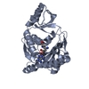
PDBj- Assembly
| Deposited unit | 
| ||||||||
|---|---|---|---|---|---|---|---|---|---|
| 1 |
| ||||||||
| Unit cell |
| ||||||||
| Components on special symmetry positions |
| ||||||||
| Details | AUTHORS STATE THAT THE ASSEMBLY OF THE BIOLOGICAL UNIT THAT IS SHOWN IN REMARK 350 IS PUTATIVE AT THE TIME OF DEPOSITION. |
-Components
| #1: Protein | Mass: 30539.324 Da / Num. of mol.: 1 Source method: isolated from a genetically manipulated source Source: (gene. exp.) Pyrococcus horikoshii (archaea) / Strain: OT3 / Gene: PH0321 / Plasmid: modified pET26 / Species (production host): Escherichia coli / Production host:  Escherichia coli BL21(DE3) (bacteria) / Strain (production host): BL21(DE3) / References: UniProt: O58059 Escherichia coli BL21(DE3) (bacteria) / Strain (production host): BL21(DE3) / References: UniProt: O58059 |
|---|---|
| #2: Protein/peptide | Mass: 392.450 Da / Num. of mol.: 1 / Source method: obtained synthetically / Details: Unknown peptide |
| #3: Water | ChemComp-HOH / Water Mass: 18.015 Da / Num. of mol.: 192 / Source method: isolated from a natural source / Formula: H2O Mass: 18.015 Da / Num. of mol.: 192 / Source method: isolated from a natural source / Formula: H2O |
-Experimental details
-Experiment
| Experiment | Method: X-RAY DIFFRACTION / Number of used crystals: 1 |
|---|
- Sample preparation
| Crystal | Density Matthews: 2.77 Å3/Da / Density % sol: 55.56 % |
|---|---|
| Crystal grow | Temperature: 294 K / Method: vapor diffusion / pH: 7 Details: 100mM Tri-sodium citrate dihydrate pH 7.0, 20% PEG 4000, 20% Isopropanol, VAPOR DIFFUSION, temperature 294K |
-Data collection
| Diffraction | Mean temperature: 100 K |
|---|---|
| Diffraction source | Source: SYNCHROTRON / Site: APS  / Beamline: 31-ID / Wavelength: 0.97958 Å / Beamline: 31-ID / Wavelength: 0.97958 Å |
| Detector | Type: MAR CCD 165 mm / Detector: CCD / Date: Dec 9, 2007 |
| Radiation | Monochromator: Diamond / Protocol: SINGLE WAVELENGTH / Monochromatic (M) / Laue (L): M / Scattering type: x-ray |
| Radiation wavelength | Wavelength: 0.97958 Å / Relative weight: 1 |
| Reflection | Resolution: 1.6→57.378 Å / Num. all: 43226 / Num. obs: 43226 / % possible obs: 97.17 % / Observed criterion σ(F): 0 / Observed criterion σ(I): 0 / Redundancy: 7.6 % / Biso Wilson estimate: 19.5 Å2 / Rmerge(I) obs: 0.095 / Rsym value: 0.095 / Net I/σ(I): 13.5 |
| Reflection shell | Resolution: 1.6→1.69 Å / Redundancy: 7.5 % / Rmerge(I) obs: 0.669 / Mean I/σ(I) obs: 2.8 / Num. unique all: 6216 / Rsym value: 0.669 / % possible all: 96.5 |
- Processing
| Software |
| ||||||||||||||||||||||||||||||||||||||||||||||||||||||||||||||||||||||||||||||||||||||||||
|---|---|---|---|---|---|---|---|---|---|---|---|---|---|---|---|---|---|---|---|---|---|---|---|---|---|---|---|---|---|---|---|---|---|---|---|---|---|---|---|---|---|---|---|---|---|---|---|---|---|---|---|---|---|---|---|---|---|---|---|---|---|---|---|---|---|---|---|---|---|---|---|---|---|---|---|---|---|---|---|---|---|---|---|---|---|---|---|---|---|---|---|
| Refinement | Method to determine structure: SAD / Resolution: 1.6→20 Å / Cor.coef. Fo:Fc: 0.96 / Cor.coef. Fo:Fc free: 0.946 / SU B: 1.426 / SU ML: 0.051 / Cross valid method: THROUGHOUT / σ(F): 0 / ESU R: 0.081 / ESU R Free: 0.081 / Stereochemistry target values: MAXIMUM LIKELIHOOD / Details: HYDROGENS HAVE BEEN ADDED IN THE RIDING POSITIONS
| ||||||||||||||||||||||||||||||||||||||||||||||||||||||||||||||||||||||||||||||||||||||||||
| Solvent computation | Ion probe radii: 0.8 Å / Shrinkage radii: 0.8 Å / VDW probe radii: 1.2 Å / Solvent model: BABINET MODEL WITH MASK | ||||||||||||||||||||||||||||||||||||||||||||||||||||||||||||||||||||||||||||||||||||||||||
| Displacement parameters | Biso mean: 24.401 Å2
| ||||||||||||||||||||||||||||||||||||||||||||||||||||||||||||||||||||||||||||||||||||||||||
| Refinement step | Cycle: LAST / Resolution: 1.6→20 Å
| ||||||||||||||||||||||||||||||||||||||||||||||||||||||||||||||||||||||||||||||||||||||||||
| Refine LS restraints |
| ||||||||||||||||||||||||||||||||||||||||||||||||||||||||||||||||||||||||||||||||||||||||||
| LS refinement shell | Resolution: 1.6→1.641 Å / Total num. of bins used: 20
|
