Resolution: 1.175→25.318 Å / SU ML: 0.13 / σ(F): 1.34 / Phase error: 14.24 / Stereochemistry target values: ML Details: ASN101 LIES BEYOND THE REASONABLE RANGE OF RAMACHANDRAN PLOT DUE TO ITS INTERACTION WITH THE SURROUNDING RESIDUES. THUS, ASN101 IS LIMITED IN A RIGID CONDITION.
Rfactor
Num. reflection
% reflection
Rfree
0.1693
1809
5 %
Rwork
0.1564
-
-
obs
0.1571
36101
99 %
Solvent computation
Shrinkage radii: 0.9 Å / VDW probe radii: 1.11 Å / Solvent model: FLAT BULK SOLVENT MODEL / Bsol: 31.88 Å2 / ksol: 0.37 e/Å3
Displacement parameters
Biso mean: 10.24 Å2
Baniso -1
Baniso -2
Baniso -3
1-
0.0998 Å2
0 Å2
-1.4265 Å2
2-
-
-0.5234 Å2
0 Å2
3-
-
-
0.4236 Å2
Refinement step
Cycle: LAST / Resolution: 1.175→25.318 Å
Protein
Nucleic acid
Ligand
Solvent
Total
Num. atoms
826
0
34
219
1079
Refine LS restraints
Refine-ID
Type
Dev ideal
Number
X-RAY DIFFRACTION
f_bond_d
0.005
883
X-RAY DIFFRACTION
f_angle_d
1.189
1204
X-RAY DIFFRACTION
f_dihedral_angle_d
32.94
343
X-RAY DIFFRACTION
f_chiral_restr
0.082
135
X-RAY DIFFRACTION
f_plane_restr
0.005
147
LS refinement shell
Resolution (Å)
Rfactor Rfree
Num. reflection Rfree
Rfactor Rwork
Num. reflection Rwork
Refine-ID
% reflection obs (%)
1.1754-1.2174
0.1724
158
0.1566
3389
X-RAY DIFFRACTION
99
1.2174-1.2661
0.1552
163
0.1506
3427
X-RAY DIFFRACTION
100
1.2661-1.3237
0.1756
196
0.1465
3441
X-RAY DIFFRACTION
100
1.3237-1.3935
0.1602
159
0.1468
3445
X-RAY DIFFRACTION
100
1.3935-1.4808
0.1634
192
0.14
3422
X-RAY DIFFRACTION
100
1.4808-1.5951
0.1417
177
0.1352
3444
X-RAY DIFFRACTION
100
1.5951-1.7556
0.1769
180
0.1453
3466
X-RAY DIFFRACTION
100
1.7556-2.0096
0.1472
195
0.1532
3404
X-RAY DIFFRACTION
98
2.0096-2.5315
0.192
203
0.1584
3301
X-RAY DIFFRACTION
96
2.5315-25.3238
0.1641
186
0.1632
3553
X-RAY DIFFRACTION
98
+
About Yorodumi
-
News
-
Feb 9, 2022. New format data for meta-information of EMDB entries
New format data for meta-information of EMDB entries
Version 3 of the EMDB header file is now the official format.
The previous official version 1.9 will be removed from the archive.
In the structure databanks used in Yorodumi, some data are registered as the other names, "COVID-19 virus" and "2019-nCoV". Here are the details of the virus and the list of structure data.
Jan 31, 2019. EMDB accession codes are about to change! (news from PDBe EMDB page)
EMDB accession codes are about to change! (news from PDBe EMDB page)
The allocation of 4 digits for EMDB accession codes will soon come to an end. Whilst these codes will remain in use, new EMDB accession codes will include an additional digit and will expand incrementally as the available range of codes is exhausted. The current 4-digit format prefixed with “EMD-” (i.e. EMD-XXXX) will advance to a 5-digit format (i.e. EMD-XXXXX), and so on. It is currently estimated that the 4-digit codes will be depleted around Spring 2019, at which point the 5-digit format will come into force.
The EM Navigator/Yorodumi systems omit the EMD- prefix.
Related info.:Q: What is EMD? / ID/Accession-code notation in Yorodumi/EM Navigator
Yorodumi is a browser for structure data from EMDB, PDB, SASBDB, etc.
This page is also the successor to EM Navigator detail page, and also detail information page/front-end page for Omokage search.
The word "yorodu" (or yorozu) is an old Japanese word meaning "ten thousand". "mi" (miru) is to see.
Related info.:EMDB / PDB / SASBDB / Comparison of 3 databanks / Yorodumi Search / Aug 31, 2016. New EM Navigator & Yorodumi / Yorodumi Papers / Jmol/JSmol / Function and homology information / Changes in new EM Navigator and Yorodumi
 Movie
Movie Controller

 Yorodumi
 Open data
 Basic information
 Components
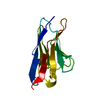
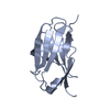
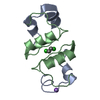
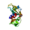
 Glucan 1,4-a-glucosidase
  Keywords
 Function and homology information
 Authors
 Citation
 Structure visualization
 Downloads & links
 Other downloads
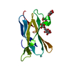


 PDBj
PDBj
 Assembly

 Mass: 18.015 Da / Num. of mol.: 219 / Source method: isolated from a natural source / Formula: H2O
 Sample preparation
 / Beamline: BL13C1 / Wavelength: 0.9762
  Processing