 Movie
Movie Controller


+ Open data

- Basic information
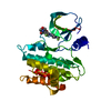
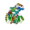
| Entry | Database: PDB / ID: 4bf7 | ||||||
|---|---|---|---|---|---|---|---|
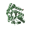
| Title | Emericilla nidulans endo-beta-1,4-galactanase | ||||||
 Components Components | ARABINOGALACTAN ENDO-1,4-BETA-GALACTOSIDASE A | ||||||
 Keywords Keywords |  HYDROLASE HYDROLASE | ||||||
| Function / homology |  Function and homology information Function and homology informationarabinogalactan endo-beta-1,4-galactanase / arabinogalactan endo-1,4-beta-galactosidase activity / glucosidase activity / pectin catabolic process / cell wall organization / extracellular regionSimilarity search - Function | ||||||
| Biological species |  Emericella nidulans (mold) Emericella nidulans (mold) | ||||||
| Method | X-RAY DIFFRACTION / SYNCHROTRON / MOLECULAR REPLACEMENT / Resolution: 2.001 Å | ||||||
 Authors Authors | Otten, H. / Michalak, M. / Larsen, S. / Mikkelsen, J.D. | ||||||
 Citation Citation | Journal: Acta Crystallogr.,Sect.F / Year: 2013 Title: The Binding of Zinc Ions to Emericella Nidulans Endo-[Beta]-1,4-Galactanase is Essential for Crystal Formation Authors: Otten, H. / Michalak, M. / Mikkelsen, J.D. / Larsen, S. | ||||||
| History |
| ||||||
| Remark 700 | SHEET DETERMINATION METHOD: DSSP THE SHEETS PRESENTED AS "AA" IN EACH CHAIN ON SHEET RECORDS BELOW ... SHEET DETERMINATION METHOD: DSSP THE SHEETS PRESENTED AS "AA" IN EACH CHAIN ON SHEET RECORDS BELOW IS ACTUALLY AN 8-STRANDED BARREL THIS IS REPRESENTED BY A 9-STRANDED SHEET IN WHICH THE FIRST AND LAST STRANDS ARE IDENTICAL. |
- Structure visualization
| Structure viewer | Molecule: MolmilJmol/JSmol |
|---|
- Downloads & links
-Download
| PDBx/mmCIF format | 4bf7.cif.gz | 142.2 KB | Display | PDBx/mmCIF format |
|---|---|---|---|---|
| PDB format | pdb4bf7.ent.gz | 111.7 KB | Display | PDB format |
| PDBx/mmJSON format | 4bf7.json.gz | Tree view | PDBx/mmJSON format | |
| Others |  Other downloads Other downloads |
-Validation report
| Arichive directory | https://data.pdbj.org/pub/pdb/validation_reports/bf/4bf7ftp://data.pdbj.org/pub/pdb/validation_reports/bf/4bf7 | HTTPS FTP |
|---|
-Related structure data
| Related structure data | 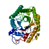 1fobS S: Starting model for refinement |
|---|---|
| Similar structure data |


-Links
 PDBj
PDBj


- Assembly
| Deposited unit | 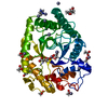
| ||||||||
|---|---|---|---|---|---|---|---|---|---|
| 1 |
| ||||||||
| Unit cell |
|
-Components
-Protein / Sugars , 2 types, 2 molecules A
| #1: Protein | Mass: 38736.637 Da / Num. of mol.: 1 Source method: isolated from a genetically manipulated source Details: SULFIDE BRIDGES BETWEEN A C253-C310 Source: (gene. exp.) Emericella nidulans (strain FGSC A4 / ATCC 38163 / CBS 112.46 / NRRL 194 / M139) (mold)Production host: KOMAGATAELLA PASTORIS (fungus)References: UniProt: Q5B153, arabinogalactan endo-beta-1,4-galactanase |
|---|---|
| #3: Sugar | ChemComp-NAG / N-Acetylglucosamine Type: D-saccharide, beta linking / Mass: 221.208 Da / Num. of mol.: 1 Type: D-saccharide, beta linking / Mass: 221.208 Da / Num. of mol.: 1Source method: isolated from a genetically manipulated source Formula: C8H15NO6 |
-Non-polymers , 5 types, 209 molecules 


| #2: Chemical | ChemComp-ZN /  Mass: 65.409 Da / Num. of mol.: 15 / Source method: obtained synthetically / Formula: Zn Mass: 65.409 Da / Num. of mol.: 15 / Source method: obtained synthetically / Formula: Zn#4: Chemical | ChemComp-ACT / Acetate Mass: 59.044 Da / Num. of mol.: 11 / Source method: obtained synthetically / Formula: C2H3O2 Mass: 59.044 Da / Num. of mol.: 11 / Source method: obtained synthetically / Formula: C2H3O2#5: Chemical | ChemComp-IMD / | Imidazole Mass: 69.085 Da / Num. of mol.: 1 / Source method: obtained synthetically / Formula: C3H5N2 Mass: 69.085 Da / Num. of mol.: 1 / Source method: obtained synthetically / Formula: C3H5N2#6: Chemical | ChemComp-GOL / | Glycerol Mass: 92.094 Da / Num. of mol.: 1 / Source method: obtained synthetically / Formula: C3H8O3 Mass: 92.094 Da / Num. of mol.: 1 / Source method: obtained synthetically / Formula: C3H8O3#7: Water | ChemComp-HOH / | WaterMass: 18.015 Da / Num. of mol.: 181 / Source method: isolated from a natural source / Formula: H2O |
|---|
-Details
| Sequence details | FUNGAL GENETICS STOCK CENTER |
|---|
-Experimental details
-Experiment
| Experiment | Method: X-RAY DIFFRACTION / Number of used crystals: 1 |
|---|
- Sample preparation
| Crystal | Density Matthews: 3 Å3/Da / Density % sol: 59.5 % / Description: NONE |
|---|---|
| Crystal grow | pH: 8 Details: 200 MM ZN ACETATE, 100 MM IMIDAZOLE, 20%(W/V) PEG 3000, PH 8.0 |
-Data collection
| Diffraction | Mean temperature: 100 K |
|---|---|
| Diffraction source | Source: SYNCHROTRON / Site: ESRF  / Beamline: ID23-2 / Wavelength: 0.8726 / Beamline: ID23-2 / Wavelength: 0.8726 |
| Detector | Type: MARRESEARCH / Detector: CCD / Date: Nov 14, 2012 Details: ONE PAIR OF PT COATED SI FOCUSING MIRRORS, KIRKPATRICK-BAEZ GEOMETRY |
| Radiation | Monochromator: HORIZONTALLY DIFFRACTING SI (111) / Protocol: SINGLE WAVELENGTH / Monochromatic (M) / Laue (L): M / Scattering type: x-ray |
| Radiation wavelength | Wavelength: 0.8726 Å / Relative weight: 1 |
| Reflection | Resolution: 2→47 Å / Num. obs: 58261 / % possible obs: 99.4 % / Observed criterion σ(I): 2 / Redundancy: 3.9 % / Biso Wilson estimate: 20.88 Å2 / Rmerge(I) obs: 0.14 / Net I/σ(I): 9.3 |
| Reflection shell | Resolution: 2→2.12 Å / Redundancy: 3.8 % / Rmerge(I) obs: 0.77 / Mean I/σ(I) obs: 2 / % possible all: 96.5 |
- Processing
| Software |
| ||||||||||||||||||||||||||||||||||||||||||||||||||||||||||||||||||||||||||||||||||||||||||||||||||||||||||||||||||||||||||||||||||||||||||||||||||||||||||
|---|---|---|---|---|---|---|---|---|---|---|---|---|---|---|---|---|---|---|---|---|---|---|---|---|---|---|---|---|---|---|---|---|---|---|---|---|---|---|---|---|---|---|---|---|---|---|---|---|---|---|---|---|---|---|---|---|---|---|---|---|---|---|---|---|---|---|---|---|---|---|---|---|---|---|---|---|---|---|---|---|---|---|---|---|---|---|---|---|---|---|---|---|---|---|---|---|---|---|---|---|---|---|---|---|---|---|---|---|---|---|---|---|---|---|---|---|---|---|---|---|---|---|---|---|---|---|---|---|---|---|---|---|---|---|---|---|---|---|---|---|---|---|---|---|---|---|---|---|---|---|---|---|---|---|---|
| Refinement | Method to determine structure: MOLECULAR REPLACEMENT Starting model: PDB ENTRY 1FOB Resolution: 2.001→47.475 Å / SU ML: 0.18 / σ(F): 1.21 / Phase error: 17.04 / Stereochemistry target values: ML Details: N-TERMINAL 1-17 MILSSLLPLSLVTLTSA IS DISORDERED AND NOT MODELLED
| ||||||||||||||||||||||||||||||||||||||||||||||||||||||||||||||||||||||||||||||||||||||||||||||||||||||||||||||||||||||||||||||||||||||||||||||||||||||||||
| Solvent computation | Shrinkage radii: 0.9 Å / VDW probe radii: 1.11 Å / Solvent model: FLAT BULK SOLVENT MODEL | ||||||||||||||||||||||||||||||||||||||||||||||||||||||||||||||||||||||||||||||||||||||||||||||||||||||||||||||||||||||||||||||||||||||||||||||||||||||||||
| Displacement parameters | Biso mean: 26.87 Å2
| ||||||||||||||||||||||||||||||||||||||||||||||||||||||||||||||||||||||||||||||||||||||||||||||||||||||||||||||||||||||||||||||||||||||||||||||||||||||||||
| Refinement step | Cycle: LAST / Resolution: 2.001→47.475 Å
| ||||||||||||||||||||||||||||||||||||||||||||||||||||||||||||||||||||||||||||||||||||||||||||||||||||||||||||||||||||||||||||||||||||||||||||||||||||||||||
| Refine LS restraints |
| ||||||||||||||||||||||||||||||||||||||||||||||||||||||||||||||||||||||||||||||||||||||||||||||||||||||||||||||||||||||||||||||||||||||||||||||||||||||||||
| LS refinement shell |
|
