 Movie
Movie Controller


+ Open data

- Basic information
| Entry | Database: PDB / ID: 4b8l | ||||||
|---|---|---|---|---|---|---|---|
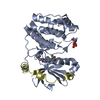
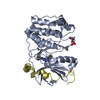
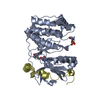
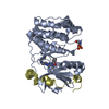
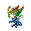
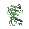
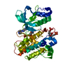
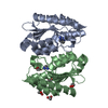
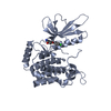
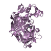
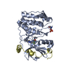
| Title | Aurora B kinase P353G mutant | ||||||
 Components Components |
| ||||||
 Keywords Keywords |  CELL CYCLE / CANCER CELL CYCLE / CANCER | ||||||
| Function / homology |  Function and homology information Function and homology informationmeiotic spindle midzone / mitotic cytokinesis checkpoint signaling / negative regulation of cytokinesis / positive regulation of mitotic sister chromatid segregation / meiotic spindle midzone assembly / metaphase chromosome alignment / abscission / mitotic spindle midzone assembly / cleavage furrow formation / spindle pole centrosome ...meiotic spindle midzone / mitotic cytokinesis checkpoint signaling / negative regulation of cytokinesis / positive regulation of mitotic sister chromatid segregation / meiotic spindle midzone assembly / metaphase chromosome alignment / abscission / mitotic spindle midzone assembly / cleavage furrow formation / spindle pole centrosome / histone H3S10 kinase activity / chromosome passenger complex / protein localization to kinetochore / negative regulation of B cell apoptotic process / positive regulation of cytokinesis / chromosome, centromeric region / mitotic cytokinesis / spindle assembly / spindle midzone / pericentric heterochromatin / post-translational protein modification / chromosome segregation / spindle microtubule / kinetochore / spindle / cellular response to UV / chromosome / midbody / microtubule / non-specific serine/threonine protein kinase / protein phosphorylation / protein serine kinase activity / protein serine/threonine kinase activity / chromatin / protein kinase binding / negative regulation of transcription by RNA polymerase II / ATP binding / metal ion binding / nucleus / cytoplasmSimilarity search - Function | ||||||
| Biological species | XENOPUS LAEVIS (African clawed frog) | ||||||
| Method | X-RAY DIFFRACTION / SYNCHROTRON / MOLECULAR REPLACEMENT / Resolution: 3 Å | ||||||
 Authors Authors | Sessa, F. / Villa, F. | ||||||
 Citation Citation | Journal: To be Published Title: Structural and Biochemical Analysis of an Aurora B Kinase Mutant Reveals a Multistep Activation Mechanism Authors: Sessa, F. / Villa, F. | ||||||
| History |
|
- Structure visualization
| Structure viewer | Molecule: MolmilJmol/JSmol |
|---|
- Downloads & links
-Download
| PDBx/mmCIF format | 4b8l.cif.gz | 73.5 KB | Display | PDBx/mmCIF format |
|---|---|---|---|---|
| PDB format | pdb4b8l.ent.gz | 54.1 KB | Display | PDB format |
| PDBx/mmJSON format | 4b8l.json.gz | Tree view | PDBx/mmJSON format | |
| Others |  Other downloads Other downloads |
-Validation report
| Arichive directory | https://data.pdbj.org/pub/pdb/validation_reports/b8/4b8lftp://data.pdbj.org/pub/pdb/validation_reports/b8/4b8l | HTTPS FTP |
|---|
-Related structure data


-Links
 PDBj
PDBj
- Assembly
| Deposited unit | 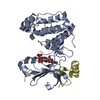
| ||||||||
|---|---|---|---|---|---|---|---|---|---|
| 1 |
| ||||||||
| Unit cell |
|
-Components
| #1: Protein | Mass: 33603.992 Da / Num. of mol.: 1 / Fragment: RESIDUES 78-361 / Mutation: YES Source method: isolated from a genetically manipulated source Source: (gene. exp.) XENOPUS LAEVIS (African clawed frog) / Production host:  ESCHERICHIA COLI (E. coli) ESCHERICHIA COLI (E. coli)References: UniProt: Q6DE08, non-specific serine/threonine protein kinase |
|---|---|
| #2: Protein/peptide | Mass: 5073.755 Da / Num. of mol.: 1 / Fragment: RESIDUES 797-840 Source method: isolated from a genetically manipulated source Source: (gene. exp.) XENOPUS LAEVIS (African clawed frog) / Production host: ESCHERICHIA COLI (E. coli) / References: UniProt: O13024 |
| #3: Chemical | ChemComp-A0P /   Mass: 506.196 Da / Num. of mol.: 1 / Source method: obtained synthetically / Formula: C10H17N6O12P3 Mass: 506.196 Da / Num. of mol.: 1 / Source method: obtained synthetically / Formula: C10H17N6O12P3 |
| Nonpolymer details | THE LIGAND HAS BEEN DEPOSITED UNDER THE HETEROGEN NAME A0P SEE REMARK 3 FOR DETAILS ON REFINEMENT ...THE LIGAND HAS BEEN DEPOSITED UNDER THE HETEROGEN NAME A0P SEE REMARK 3 FOR DETAILS ON REFINEMENT |
-Experimental details
-Experiment
| Experiment | Method: X-RAY DIFFRACTION |
|---|
- Sample preparation
| Crystal | Density Matthews: 2.34 Å3/Da / Density % sol: 47.39 % / Description: NONE |
|---|
-Data collection
| Diffraction | Mean temperature: 100 K |
|---|---|
| Diffraction source | Source: SYNCHROTRON / Site: ESRF  / Beamline: ID14-3 / Wavelength: 0.934 / Beamline: ID14-3 / Wavelength: 0.934 |
| Detector | Type: ADSC CCD / Detector: CCD / Date: Jun 18, 2006 |
| Radiation | Protocol: SINGLE WAVELENGTH / Monochromatic (M) / Laue (L): M / Scattering type: x-ray |
| Radiation wavelength | Wavelength: 0.934 Å / Relative weight: 1 |
| Reflection | Resolution: 3→25 Å / Num. obs: 7248 / % possible obs: 99 % / Observed criterion σ(I): 2 / Redundancy: 3.5 % / Rmerge(I) obs: 0.05 / Net I/σ(I): 19.5 |
| Reflection shell | Resolution: 3→3.09 Å / Redundancy: 3.5 % / Rmerge(I) obs: 0.35 / Mean I/σ(I) obs: 3.8 / % possible all: 95.4 |
- Processing
| Software |
| ||||||||||||||||||||||||||||||||||||||||||||||||||||||||||||||||||||||||||||||||||||||||||||||||||||||||||||||||||||||||||||||||||||||||||||||||||||||||||||||||||||||||||||||||||||||
|---|---|---|---|---|---|---|---|---|---|---|---|---|---|---|---|---|---|---|---|---|---|---|---|---|---|---|---|---|---|---|---|---|---|---|---|---|---|---|---|---|---|---|---|---|---|---|---|---|---|---|---|---|---|---|---|---|---|---|---|---|---|---|---|---|---|---|---|---|---|---|---|---|---|---|---|---|---|---|---|---|---|---|---|---|---|---|---|---|---|---|---|---|---|---|---|---|---|---|---|---|---|---|---|---|---|---|---|---|---|---|---|---|---|---|---|---|---|---|---|---|---|---|---|---|---|---|---|---|---|---|---|---|---|---|---|---|---|---|---|---|---|---|---|---|---|---|---|---|---|---|---|---|---|---|---|---|---|---|---|---|---|---|---|---|---|---|---|---|---|---|---|---|---|---|---|---|---|---|---|---|---|---|---|
| Refinement | Method to determine structure: MOLECULAR REPLACEMENT / Resolution: 3→50 Å / Cor.coef. Fo:Fc: 0.914 / Cor.coef. Fo:Fc free: 0.87 / SU B: 24.42 / SU ML: 0.436 / Cross valid method: THROUGHOUT / ESU R Free: 0.529 / Stereochemistry target values: MAXIMUM LIKELIHOOD Details: HYDROGENS HAVE BEEN ADDED IN THE RIDING POSITIONS. THE AUTHORS NOTICED DURING THE REFINEMENT OF THE A0P LIGAND A DIFFERENCE IN THE CANONICAL CHIRALITY DUE TO THE TORSION OF THE LIGAND ITSELF ...Details: HYDROGENS HAVE BEEN ADDED IN THE RIDING POSITIONS. THE AUTHORS NOTICED DURING THE REFINEMENT OF THE A0P LIGAND A DIFFERENCE IN THE CANONICAL CHIRALITY DUE TO THE TORSION OF THE LIGAND ITSELF WITHIN THE ACTIVE SITE OF THE ENZYME, AND THEREFORE DECIDED TO DEPOSIT THE COORDINATES AS THEY APPEAR IN THIS ENTRY.
| ||||||||||||||||||||||||||||||||||||||||||||||||||||||||||||||||||||||||||||||||||||||||||||||||||||||||||||||||||||||||||||||||||||||||||||||||||||||||||||||||||||||||||||||||||||||
| Solvent computation | Ion probe radii: 0.8 Å / Shrinkage radii: 0.8 Å / VDW probe radii: 1.2 Å / Solvent model: MASK | ||||||||||||||||||||||||||||||||||||||||||||||||||||||||||||||||||||||||||||||||||||||||||||||||||||||||||||||||||||||||||||||||||||||||||||||||||||||||||||||||||||||||||||||||||||||
| Displacement parameters | Biso mean: 65.929 Å2
| ||||||||||||||||||||||||||||||||||||||||||||||||||||||||||||||||||||||||||||||||||||||||||||||||||||||||||||||||||||||||||||||||||||||||||||||||||||||||||||||||||||||||||||||||||||||
| Refinement step | Cycle: LAST / Resolution: 3→50 Å
| ||||||||||||||||||||||||||||||||||||||||||||||||||||||||||||||||||||||||||||||||||||||||||||||||||||||||||||||||||||||||||||||||||||||||||||||||||||||||||||||||||||||||||||||||||||||
| Refine LS restraints |
|