 Movie
Movie Controller

[English] 日本語
 Yorodumi
- PDB-4ase: CRYSTAL STRUCTURE OF VEGFR2 (JUXTAMEMBRANE AND KINASE DOMAINS) IN... -
+ Open data

- Basic information
| Entry | Database: PDB / ID: 4ase | ||||||
|---|---|---|---|---|---|---|---|
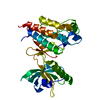
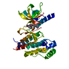
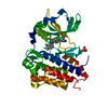
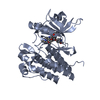
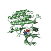
| Title | CRYSTAL STRUCTURE OF VEGFR2 (JUXTAMEMBRANE AND KINASE DOMAINS) IN COMPLEX WITH TIVOZANIB (AV-951) | ||||||
 Components Components | VASCULAR ENDOTHELIAL GROWTH FACTOR RECEPTOR 2 VEGF receptor VEGF receptor | ||||||
 Keywords Keywords | TRANSFERASE / ANGIOGENESIS / SIGNALING PROTEIN / PHOSPHORYLATION / RECEPTOR / INHIBITOR | ||||||
| Function / homology |  Function and homology information Function and homology informationblood vessel endothelial cell differentiation / cellular response to hydrogen sulfide / regulation of bone development / Signaling by membrane-tethered fusions of PDGFRA or PDGFRB / vascular endothelial growth factor binding / Neurophilin interactions with VEGF and VEGFR / endothelium development / vascular endothelial growth factor receptor-2 signaling pathway / VEGF binds to VEGFR leading to receptor dimerization / endocardium development ...blood vessel endothelial cell differentiation / cellular response to hydrogen sulfide / regulation of bone development / Signaling by membrane-tethered fusions of PDGFRA or PDGFRB / vascular endothelial growth factor binding / Neurophilin interactions with VEGF and VEGFR / endothelium development / vascular endothelial growth factor receptor-2 signaling pathway / VEGF binds to VEGFR leading to receptor dimerization / endocardium development / vascular wound healing / vascular endothelial growth factor receptor activity / regulation of hematopoietic progenitor cell differentiation / post-embryonic camera-type eye morphogenesis / endothelial cell differentiation / mesenchymal cell proliferation / positive regulation of vasculogenesis / lymph vessel development / positive regulation of BMP signaling pathway / surfactant homeostasis / cell migration involved in sprouting angiogenesis / anchoring junction / epithelial cell maturation / positive regulation of positive chemotaxis / embryonic hemopoiesis / vascular endothelial growth factor signaling pathway / positive regulation of endothelial cell chemotaxis / positive regulation of mesenchymal cell proliferation / positive regulation of mitochondrial depolarization / branching involved in blood vessel morphogenesis / positive regulation of cell migration involved in sprouting angiogenesis / positive regulation of mitochondrial fission / lung alveolus development / positive regulation of stem cell proliferation / positive regulation of nitric-oxide synthase biosynthetic process / sorting endosome / growth factor binding / positive regulation of focal adhesion assembly / regulation of MAPK cascade / semaphorin-plexin signaling pathway / positive regulation of macroautophagy / : / positive regulation of blood vessel endothelial cell migration / cellular response to vascular endothelial growth factor stimulus / cell fate commitment / calcium ion homeostasis / Integrin cell surface interactions / vasculogenesis / vascular endothelial growth factor receptor signaling pathway / coreceptor activity / negative regulation of endothelial cell apoptotic process / peptidyl-tyrosine autophosphorylation / ovarian follicle development / positive regulation of endothelial cell proliferation / transmembrane receptor protein tyrosine kinase activity / positive regulation of endothelial cell migration / VEGFR2 mediated cell proliferation / epithelial cell proliferation / stem cell proliferation / cell surface receptor protein tyrosine kinase signaling pathway / Hsp90 protein binding / receptor protein-tyrosine kinase / VEGFA-VEGFR2 Pathway / peptidyl-tyrosine phosphorylation / positive regulation of angiogenesis / cell migration / integrin binding / cell junction / regulation of cell shape / protein tyrosine kinase activity / angiogenesis / negative regulation of neuron apoptotic process / positive regulation of MAPK cascade / protein autophosphorylation / positive regulation of phosphatidylinositol 3-kinase/protein kinase B signal transduction / early endosome / positive regulation of ERK1 and ERK2 cascade / receptor complex / endosome / positive regulation of cell migration / cadherin binding / membrane raft / positive regulation of protein phosphorylation / external side of plasma membrane / negative regulation of gene expression / positive regulation of cell population proliferation / negative regulation of apoptotic process / Golgi apparatus / endoplasmic reticulum / extracellular region / ATP binding / identical protein binding / nucleus / plasma membraneSimilarity search - Function | ||||||
| Biological species |  HOMO SAPIENS (human) HOMO SAPIENS (human) | ||||||
| Method | X-RAY DIFFRACTION / SYNCHROTRON / MOLECULAR REPLACEMENT / Resolution: 1.83 Å | ||||||
 Authors Authors | McTigue, M. / Deng, Y. / Ryan, K. / Brooun, A. / Diehl, W. / Stewart, A. | ||||||
 Citation Citation | Journal: Proc.Natl.Acad.Sci.USA / Year: 2012 Title: Molecular Conformations, Interactions, and Properties Associated with Drug Efficiency and Clinical Performance Among Vegfr Tk Inhibitors. Authors: Mctigue, M. / Murray, B.W. / Chen, J.H. / Deng, Y. / Solowiej, J. / Kania, R.S. | ||||||
| History |
|
- Structure visualization
| Structure viewer | Molecule: MolmilJmol/JSmol |
|---|
- Downloads & links
-Download
| PDBx/mmCIF format | 4ase.cif.gz | 84.5 KB | Display | PDBx/mmCIF format |
|---|---|---|---|---|
| PDB format | pdb4ase.ent.gz | 60.6 KB | Display | PDB format |
| PDBx/mmJSON format | 4ase.json.gz | Tree view | PDBx/mmJSON format | |
| Others |  Other downloads Other downloads |
-Validation report
| Arichive directory | https://data.pdbj.org/pub/pdb/validation_reports/as/4aseftp://data.pdbj.org/pub/pdb/validation_reports/as/4ase | HTTPS FTP |
|---|
-Related structure data
| Related structure data | 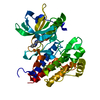 4ag8C  4agcC  4agdC 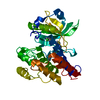 4asdSC C: citing same article ( S: Starting model for refinement |
|---|---|
| Similar structure data |


-Links
 PDBj
PDBj


- Assembly
| Deposited unit | 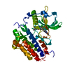
| ||||||||
|---|---|---|---|---|---|---|---|---|---|
| 1 |
| ||||||||
| Unit cell |
|
-Components
| #1: Protein | VEGF receptor / VEGFR-2 / FETAL LIVER KINASE 1 / FLK-1 / KINASE INSERT DOMAIN RECEPTOR / KDR / PROTEIN-TYROSINE ...VEGFR-2 / FETAL LIVER KINASE 1 / FLK-1 / KINASE INSERT DOMAIN RECEPTOR / KDR / PROTEIN-TYROSINE KINASE RECEPTOR FLK-1 / CD309 Mass: 40258.391 Da / Num. of mol.: 1 Fragment: JUXTAMEMBRANE AND KINASE DOMAINS, RESIDUES 787-1171 Mutation: YES Source method: isolated from a genetically manipulated source Source: (gene. exp.) HOMO SAPIENS (human) / Plasmid: PFASTBAC / Cell line (production host): SF9 / Production host:   SPODOPTERA FRUGIPERDA (fall armyworm) SPODOPTERA FRUGIPERDA (fall armyworm)References: UniProt: P35968, receptor protein-tyrosine kinase | ||
|---|---|---|---|
| #2: Chemical | ChemComp-AV9 / Tivozanib  Mass: 454.863 Da / Num. of mol.: 1 / Source method: obtained synthetically / Formula: C22H19ClN4O5 / Comment: medication, inhibitor*YM Mass: 454.863 Da / Num. of mol.: 1 / Source method: obtained synthetically / Formula: C22H19ClN4O5 / Comment: medication, inhibitor*YM | ||
| #3: Water | ChemComp-HOH / Water Mass: 18.015 Da / Num. of mol.: 249 / Source method: isolated from a natural source / Formula: H2O Mass: 18.015 Da / Num. of mol.: 249 / Source method: isolated from a natural source / Formula: H2O | ||
| Compound details | ENGINEERED| Sequence details | IN ADDITION TO THE MUTATION E990V, RESIDUES 940-989 WERE ALSO DELETED FROM THE CRYSTALLIS | |
-Experimental details
-Experiment
| Experiment | Method: X-RAY DIFFRACTION / Number of used crystals: 1 |
|---|
- Sample preparation
| Crystal | Density Matthews: 2.6 Å3/Da / Density % sol: 53 % / Description: NONE |
|---|---|
| Crystal grow | Temperature: 286 K / Method: vapor diffusion, hanging drop / pH: 6.2 Details: CRYSTALS WERE OBTAINED BY THE HANGING DROP VAPOR DIFFUSION METHOD AT 13 DEGREES C BY THE USING PRECIPITANT SOLUTIONS CONTAINING APPROXIMATELY: 0.2M SODIUM CITRATE AND 14-21% (W/V) ...Details: CRYSTALS WERE OBTAINED BY THE HANGING DROP VAPOR DIFFUSION METHOD AT 13 DEGREES C BY THE USING PRECIPITANT SOLUTIONS CONTAINING APPROXIMATELY: 0.2M SODIUM CITRATE AND 14-21% (W/V) POLYETHYLENE GLYCOL 3350., pH 6.2 |
-Data collection
| Diffraction | Mean temperature: 85 K |
|---|---|
| Diffraction source | Source: SYNCHROTRON / Site: APS  / Beamline: 17-ID / Wavelength: 1 / Beamline: 17-ID / Wavelength: 1 |
| Detector | Type: DECTRIS PILATUS 6M / Detector: PIXEL / Date: Oct 17, 2011 |
| Radiation | Protocol: SINGLE WAVELENGTH / Monochromatic (M) / Laue (L): M / Scattering type: x-ray |
| Radiation wavelength | Wavelength: 1 Å / Relative weight: 1 |
| Reflection | Resolution: 1.83→43.2 Å / Num. obs: 33988 / % possible obs: 99.1 % / Observed criterion σ(I): 1 / Redundancy: 3.3 % / Biso Wilson estimate: 26.2 Å2 / Rmerge(I) obs: 0.03 / Net I/σ(I): 17.4 |
| Reflection shell | Resolution: 1.83→1.93 Å / Redundancy: 3.4 % / Rmerge(I) obs: 0.4 / Mean I/σ(I) obs: 2.5 / % possible all: 99.8 |
- Processing
| Software |
| ||||||||||||||||||||||||||||||||||||||||||||||||||||||||||||
|---|---|---|---|---|---|---|---|---|---|---|---|---|---|---|---|---|---|---|---|---|---|---|---|---|---|---|---|---|---|---|---|---|---|---|---|---|---|---|---|---|---|---|---|---|---|---|---|---|---|---|---|---|---|---|---|---|---|---|---|---|---|
| Refinement | Method to determine structure: MOLECULAR REPLACEMENT Starting model: PDB ENTRY 4ASD Resolution: 1.83→43.32 Å / Rfactor Rfree error: 0.007 / Data cutoff high absF: 1332184.64 / Data cutoff low absF: 0 / Isotropic thermal model: RESTRAINED / Cross valid method: THROUGHOUT / σ(F): 0
| ||||||||||||||||||||||||||||||||||||||||||||||||||||||||||||
| Solvent computation | Solvent model: FLAT MODEL / Bsol: 69.3274 Å2 / ksol: 0.373306 e/Å3 | ||||||||||||||||||||||||||||||||||||||||||||||||||||||||||||
| Displacement parameters | Biso mean: 42.3 Å2
| ||||||||||||||||||||||||||||||||||||||||||||||||||||||||||||
| Refine analyze |
| ||||||||||||||||||||||||||||||||||||||||||||||||||||||||||||
| Refinement step | Cycle: LAST / Resolution: 1.83→43.32 Å
| ||||||||||||||||||||||||||||||||||||||||||||||||||||||||||||
| Refine LS restraints |
| ||||||||||||||||||||||||||||||||||||||||||||||||||||||||||||
| Refine LS restraints NCS | NCS model details: NONE | ||||||||||||||||||||||||||||||||||||||||||||||||||||||||||||
| LS refinement shell | Resolution: 1.83→1.94 Å / Rfactor Rfree error: 0.024 / Total num. of bins used: 6
| ||||||||||||||||||||||||||||||||||||||||||||||||||||||||||||
| Xplor file |
|
