 Movie
Movie Controller


+ Open data

- Basic information
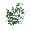
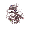
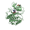
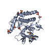
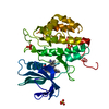
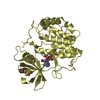
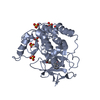
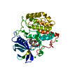
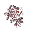
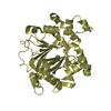
| Entry | Database: PDB / ID: 4btj | ||||||
|---|---|---|---|---|---|---|---|
| Title | TTBK1 in complex with ATP | ||||||
 Components Components | TAU-TUBULIN KINASE 1 | ||||||
 Keywords Keywords |  TRANSFERASE / LIGAND COMPLEX / STRUCTURE-KINETICS RELATIONSHIP TRANSFERASE / LIGAND COMPLEX / STRUCTURE-KINETICS RELATIONSHIP | ||||||
| Function / homology |  Function and homology information Function and homology informationpositive regulation of astrocyte activation / positive regulation of cyclin-dependent protein kinase activity / positive regulation of microglial cell activation / positive regulation of cysteine-type endopeptidase activity / microtubule associated complex / positive regulation of protein polymerization / tau-protein kinase activity / substantia nigra development / negative regulation of protein binding / peptidyl-threonine phosphorylation ...positive regulation of astrocyte activation / positive regulation of cyclin-dependent protein kinase activity / positive regulation of microglial cell activation / positive regulation of cysteine-type endopeptidase activity / microtubule associated complex / positive regulation of protein polymerization / tau-protein kinase activity / substantia nigra development / negative regulation of protein binding / peptidyl-threonine phosphorylation / tau protein binding / peptidyl-tyrosine phosphorylation / peptidyl-serine phosphorylation / protein tyrosine kinase activity / learning or memory / non-specific serine/threonine protein kinase / protein phosphorylation / negative regulation of gene expression / protein serine kinase activity / protein serine/threonine kinase activity / neuronal cell body / positive regulation of gene expression / perinuclear region of cytoplasm / signal transduction / nucleoplasm / ATP binding / nucleus / cytosolSimilarity search - Function | ||||||
| Biological species |  HOMO SAPIENS (human) HOMO SAPIENS (human) | ||||||
| Method | X-RAY DIFFRACTION / SYNCHROTRON / MOLECULAR REPLACEMENT / Resolution: 2.16 Å | ||||||
 Authors Authors | Xue, Y. / Wan, P. / Hillertz, P. / Schweikart, F. / Zhao, Y. / Wissler, L. / Dekker, N. | ||||||
 Citation Citation | Journal: Chemmedchem / Year: 2013 Title: X-Ray Structural Analysis of Tau-Tubulin Kinase 1 and its Interactions with Small Molecular Inhibitors. Authors: Xue, Y. / Wan, P.T. / Hillertz, P. / Schweikart, F. / Zhao, Y. / Wissler, L. / Dekker, N. | ||||||
| History |
|
- Structure visualization
| Structure viewer | Molecule: MolmilJmol/JSmol |
|---|
- Downloads & links
-Download
| PDBx/mmCIF format | 4btj.cif.gz | 147 KB | Display | PDBx/mmCIF format |
|---|---|---|---|---|
| PDB format | pdb4btj.ent.gz | 114.8 KB | Display | PDB format |
| PDBx/mmJSON format | 4btj.json.gz | Tree view | PDBx/mmJSON format | |
| Others |  Other downloads Other downloads |
-Validation report
| Arichive directory | https://data.pdbj.org/pub/pdb/validation_reports/bt/4btjftp://data.pdbj.org/pub/pdb/validation_reports/bt/4btj | HTTPS FTP |
|---|
-Related structure data


-Links
 PDBj
PDBj- Assembly
| Deposited unit | 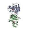
| ||||||||
|---|---|---|---|---|---|---|---|---|---|
| 1 | 
| ||||||||
| 2 | 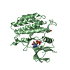
| ||||||||
| Unit cell |
|
-Components
| #1: Protein | Mass: 38746.543 Da / Num. of mol.: 2 / Fragment: KINASE DOMAIN, RESIDUES 1-313 Source method: isolated from a genetically manipulated source Source: (gene. exp.) HOMO SAPIENS (human) / Production host:   SPODOPTERA FRUGIPERDA (fall armyworm) SPODOPTERA FRUGIPERDA (fall armyworm)References: UniProt: Q5TCY1, non-specific serine/threonine protein kinase, tau-protein kinase#2: Chemical | ChemComp-ADP / | Adenosine diphosphate  Mass: 427.201 Da / Num. of mol.: 1 / Source method: obtained synthetically / Formula: C10H15N5O10P2 / Comment: ADP, energy-carrying molecule*YM Mass: 427.201 Da / Num. of mol.: 1 / Source method: obtained synthetically / Formula: C10H15N5O10P2 / Comment: ADP, energy-carrying molecule*YM#3: Chemical | ChemComp-PO4 / Phosphate  Mass: 94.971 Da / Num. of mol.: 4 / Source method: obtained synthetically / Formula: PO4 Mass: 94.971 Da / Num. of mol.: 4 / Source method: obtained synthetically / Formula: PO4#4: Chemical | ChemComp-ATP / | Adenosine triphosphate  Mass: 507.181 Da / Num. of mol.: 1 / Source method: obtained synthetically / Formula: C10H16N5O13P3 / Comment: ATP, energy-carrying molecule*YM Mass: 507.181 Da / Num. of mol.: 1 / Source method: obtained synthetically / Formula: C10H16N5O13P3 / Comment: ATP, energy-carrying molecule*YM#5: Water | ChemComp-HOH / | Water Mass: 18.015 Da / Num. of mol.: 617 / Source method: isolated from a natural source / Formula: H2O Mass: 18.015 Da / Num. of mol.: 617 / Source method: isolated from a natural source / Formula: H2O |
|---|
-Experimental details
-Experiment
| Experiment | Method: X-RAY DIFFRACTION |
|---|
- Sample preparation
| Crystal | Density Matthews: 4.93 Å3/Da / Density % sol: 78.8 % / Description: NONE |
|---|
-Data collection
| Diffraction | Mean temperature: 100 K |
|---|---|
| Diffraction source | Source: SYNCHROTRON / Site: Diamond  / Beamline: I04-1 / Wavelength: 0.9173 / Beamline: I04-1 / Wavelength: 0.9173 |
| Detector | Type: ADSC CCD / Detector: CCD |
| Radiation | Protocol: SINGLE WAVELENGTH / Monochromatic (M) / Laue (L): M / Scattering type: x-ray |
| Radiation wavelength | Wavelength: 0.9173 Å / Relative weight: 1 |
| Reflection | Resolution: 2.16→21.77 Å / Num. obs: 79065 / % possible obs: 98.1 % / Observed criterion σ(I): 1 / Redundancy: 3.4 % / Biso Wilson estimate: 36.67 Å2 / Rmerge(I) obs: 0.07 / Net I/σ(I): 11.3 |
| Reflection shell | Resolution: 2.16→2.22 Å / Redundancy: 3 % / Rmerge(I) obs: 0.52 / Mean I/σ(I) obs: 2.1 / % possible all: 87.3 |
- Processing
| Software |
| ||||||||||||||||||||||||||||||||||||||||||||||||||||||||||||||||||||||||||||||||||||||||||||||||||||||||||||||||||
|---|---|---|---|---|---|---|---|---|---|---|---|---|---|---|---|---|---|---|---|---|---|---|---|---|---|---|---|---|---|---|---|---|---|---|---|---|---|---|---|---|---|---|---|---|---|---|---|---|---|---|---|---|---|---|---|---|---|---|---|---|---|---|---|---|---|---|---|---|---|---|---|---|---|---|---|---|---|---|---|---|---|---|---|---|---|---|---|---|---|---|---|---|---|---|---|---|---|---|---|---|---|---|---|---|---|---|---|---|---|---|---|---|---|---|---|
| Refinement | Method to determine structure: MOLECULAR REPLACEMENT / Resolution: 2.16→21.77 Å / Cor.coef. Fo:Fc: 0.9461 / Cor.coef. Fo:Fc free: 0.9366 / SU R Cruickshank DPI: 0.119 / Cross valid method: THROUGHOUT / σ(F): 0 / SU R Blow DPI: 0.124 / SU Rfree Blow DPI: 0.115 / SU Rfree Cruickshank DPI: 0.112
| ||||||||||||||||||||||||||||||||||||||||||||||||||||||||||||||||||||||||||||||||||||||||||||||||||||||||||||||||||
| Displacement parameters | Biso mean: 39.27 Å2
| ||||||||||||||||||||||||||||||||||||||||||||||||||||||||||||||||||||||||||||||||||||||||||||||||||||||||||||||||||
| Refine analyze | Luzzati coordinate error obs: 0.233 Å | ||||||||||||||||||||||||||||||||||||||||||||||||||||||||||||||||||||||||||||||||||||||||||||||||||||||||||||||||||
| Refinement step | Cycle: LAST / Resolution: 2.16→21.77 Å
| ||||||||||||||||||||||||||||||||||||||||||||||||||||||||||||||||||||||||||||||||||||||||||||||||||||||||||||||||||
| Refine LS restraints |
| ||||||||||||||||||||||||||||||||||||||||||||||||||||||||||||||||||||||||||||||||||||||||||||||||||||||||||||||||||
| LS refinement shell | Resolution: 2.16→2.22 Å / Total num. of bins used: 20
|
