DIPEPTIDYLPEPTIDASE4SOLUBLEFORM / DIPEPTIDYL PEPTIDASE IV SOLUBLE FORM / DIPEPTIDYL PEPTIDASE 4 / ADABP / ADENOSINE DEAMINASE ...DIPEPTIDYL PEPTIDASE IV SOLUBLE FORM / DIPEPTIDYL PEPTIDASE 4 / ADABP / ADENOSINE DEAMINASE COMPLEXING PROTEIN 2 / ADCP-2 / DIPEPTIDYL PEPTIDASE IV / DPP IV / T-CELL ACTIVATION ANTIGEN CD26 / TP103 / CD_ANTIGEN=CD26
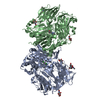
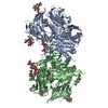
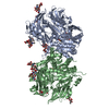
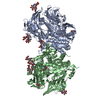
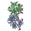
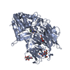
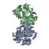
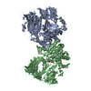
Mass: 85831.156 Da / Num. of mol.: 2 Source method: isolated from a genetically manipulated source Source: (gene. exp.) HOMO SAPIENS (human) / Production host: SPODOPTERA FRUGIPERDA (fall armyworm) / References: UniProt: P27487, dipeptidyl-peptidase IV
Mass: 18.015 Da / Num. of mol.: 1590 / Source method: isolated from a natural source / Formula: H2O
-
Details
Sequence details
S437I SEQUENCE CONFLICT KNOWN IN UNIPROT.
-
Experimental details
-
Experiment
Experiment
Method: X-RAY DIFFRACTION / Number of used crystals: 1
-
Sample preparation
Crystal
Density Matthews: 3.28 Å3/Da / Density % sol: 58.2 % / Description: NONE
Crystal grow
Details: 2 UL PROTEIN, 0.4 UL RESERVOIR, 0.25 UL WATER; PROTEIN SOLUTION: 5.2 MG/ML DPP4, 25 MM TRIS PH 8, 25 MM NACL, 2 MM NVP-BIV988-AA-1, 2% DMSO; RESERVOIR SOLUTION: 40% PEG 1000, 200 MM TRIS PH ...Details: 2 UL PROTEIN, 0.4 UL RESERVOIR, 0.25 UL WATER; PROTEIN SOLUTION: 5.2 MG/ML DPP4, 25 MM TRIS PH 8, 25 MM NACL, 2 MM NVP-BIV988-AA-1, 2% DMSO; RESERVOIR SOLUTION: 40% PEG 1000, 200 MM TRIS PH 9.0, 200 MM AMMONIUM SULFATE, 5% GLYCEROL
Type: MARRESEARCH / Detector: CCD / Date: Dec 1, 2005
Radiation
Protocol: SINGLE WAVELENGTH / Monochromatic (M) / Laue (L): M / Scattering type: x-ray
Radiation wavelength
Wavelength: 1.00054 Å / Relative weight: 1
Reflection
Resolution: 1.62→87 Å / Num. obs: 284276 / % possible obs: 99.7 % / Observed criterion σ(I): -3 / Redundancy: 7.89 % / Biso Wilson estimate: 21.73 Å2 / Rmerge(I) obs: 0.07 / Net I/σ(I): 17.22
Reflection shell
Resolution: 1.62→1.66 Å / Redundancy: 5.09 % / Rmerge(I) obs: 0.4 / Mean I/σ(I) obs: 4.2 / % possible all: 99.8
-
Processing
Software
Name
Version
Classification
BUSTER
2.11.2
refinement
XDS
datareduction
XSCALE
datascaling
Refinement
Method to determine structure: MOLECULAR REPLACEMENT / Resolution: 1.62→68.06 Å / Cor.coef. Fo:Fc: 0.9644 / Cor.coef. Fo:Fc free: 0.9596 / SU R Cruickshank DPI: 0.066 / Cross valid method: THROUGHOUT / σ(F): 0 / SU R Blow DPI: 0.068 / SU Rfree Blow DPI: 0.066 / SU Rfree Cruickshank DPI: 0.064 Details: IDEAL-DIST CONTACT TERM CONTACT SETUP. ALL ATOMS HAVE CCP4 ATOM TYPE FROM LIBRARY.
Rfactor
Num. reflection
% reflection
Selection details
Rfree
0.1789
5116
1.8 %
RANDOM
Rwork
0.1638
-
-
-
obs
0.1641
284274
99.74 %
-
Displacement parameters
Biso mean: 25.12 Å2
Baniso -1
Baniso -2
Baniso -3
1-
-0.1534 Å2
0 Å2
0 Å2
2-
-
0.2328 Å2
0 Å2
3-
-
-
-0.0794 Å2
Refine analyze
Luzzati coordinate error obs: 0.155 Å
Refinement step
Cycle: LAST / Resolution: 1.62→68.06 Å
Protein
Nucleic acid
Ligand
Solvent
Total
Num. atoms
11961
0
333
1590
13884
Refine LS restraints
Refine-ID
Type
Dev ideal
Number
Restraint function
Weight
X-RAY DIFFRACTION
t_bond_d
0.01
12792
HARMONIC
2
X-RAY DIFFRACTION
t_angle_deg
1.05
17481
HARMONIC
2
X-RAY DIFFRACTION
t_dihedral_angle_d
4368
SINUSOIDAL
2
X-RAY DIFFRACTION
t_incorr_chiral_ct
X-RAY DIFFRACTION
t_pseud_angle
X-RAY DIFFRACTION
t_trig_c_planes
311
HARMONIC
2
X-RAY DIFFRACTION
t_gen_planes
1868
HARMONIC
5
X-RAY DIFFRACTION
t_it
12792
HARMONIC
20
X-RAY DIFFRACTION
t_nbd
X-RAY DIFFRACTION
t_omega_torsion
4.3
X-RAY DIFFRACTION
t_other_torsion
17.91
X-RAY DIFFRACTION
t_improper_torsion
X-RAY DIFFRACTION
t_chiral_improper_torsion
1677
SEMIHARMONIC
5
X-RAY DIFFRACTION
t_sum_occupancies
X-RAY DIFFRACTION
t_utility_distance
X-RAY DIFFRACTION
t_utility_angle
X-RAY DIFFRACTION
t_utility_torsion
X-RAY DIFFRACTION
t_ideal_dist_contact
16099
SEMIHARMONIC
4
LS refinement shell
Resolution: 1.62→1.66 Å / Total num. of bins used: 20
Rfactor
Num. reflection
% reflection
Rfree
0.1959
374
1.8 %
Rwork
0.1924
20403
-
all
0.1925
20777
-
obs
-
-
99.74 %
+
About Yorodumi
-
News
-
Feb 9, 2022. New format data for meta-information of EMDB entries
New format data for meta-information of EMDB entries
Version 3 of the EMDB header file is now the official format.
The previous official version 1.9 will be removed from the archive.
In the structure databanks used in Yorodumi, some data are registered as the other names, "COVID-19 virus" and "2019-nCoV". Here are the details of the virus and the list of structure data.
Jan 31, 2019. EMDB accession codes are about to change! (news from PDBe EMDB page)
EMDB accession codes are about to change! (news from PDBe EMDB page)
The allocation of 4 digits for EMDB accession codes will soon come to an end. Whilst these codes will remain in use, new EMDB accession codes will include an additional digit and will expand incrementally as the available range of codes is exhausted. The current 4-digit format prefixed with “EMD-” (i.e. EMD-XXXX) will advance to a 5-digit format (i.e. EMD-XXXXX), and so on. It is currently estimated that the 4-digit codes will be depleted around Spring 2019, at which point the 5-digit format will come into force.
The EM Navigator/Yorodumi systems omit the EMD- prefix.
Related info.:Q: What is EMD? / ID/Accession-code notation in Yorodumi/EM Navigator
Yorodumi is a browser for structure data from EMDB, PDB, SASBDB, etc.
This page is also the successor to EM Navigator detail page, and also detail information page/front-end page for Omokage search.
The word "yorodu" (or yorozu) is an old Japanese word meaning "ten thousand". "mi" (miru) is to see.
Related info.:EMDB / PDB / SASBDB / Comparison of 3 databanks / Yorodumi Search / Aug 31, 2016. New EM Navigator & Yorodumi / Yorodumi Papers / Jmol/JSmol / Function and homology information / Changes in new EM Navigator and Yorodumi
 Movie
Movie Controller

 Yorodumi
 Open data
 Basic information
 Components
 Keywords
 HYDROLASE /
  Function and homology information
 Authors
 Citation
 Structure visualization
 Downloads & links
 Other downloads


 PDBj
PDBj


 Assembly


 Type: D-saccharide, beta linking / Mass: 221.208 Da / Num. of mol.: 12


 Mass: 489.571 Da / Num. of mol.: 2 / Source method: obtained synthetically / Formula: C29H27N7O
 Mass: 96.063 Da / Num. of mol.: 6 / Source method: obtained synthetically / Formula: SO4
 Sample preparation
 / Beamline: X10SA / Wavelength: 1.00054
  Processing