 Movie
Movie Controller

[English] 日本語
 Yorodumi
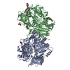
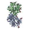
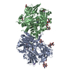
- PDB-1nu8: Crystal structure of human dipeptidyl peptidase IV (DPP-IV) in co... -
+ Open data

- Basic information
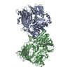
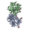
| Entry | Database: PDB / ID: 1nu8 | ||||||
|---|---|---|---|---|---|---|---|
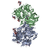
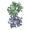
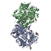
| Title | Crystal structure of human dipeptidyl peptidase IV (DPP-IV) in complex with Diprotin A (IPI) | ||||||
 Components Components |
| ||||||
 Keywords Keywords |  HYDROLASE / b-barrel / alpha/beta hydrolase fold / exopeptidase / diprotin A HYDROLASE / b-barrel / alpha/beta hydrolase fold / exopeptidase / diprotin A | ||||||
| Function / homology |  Function and homology information Function and homology informationglucagon processing / negative regulation of neutrophil chemotaxis / Synthesis, secretion, and inactivation of Glucose-dependent Insulinotropic Polypeptide (GIP) / regulation of cell-cell adhesion mediated by integrin / negative regulation of extracellular matrix disassembly / psychomotor behavior / dipeptidyl-peptidase IV / intercellular canaliculus / chemorepellent activity / dipeptidyl-peptidase activity ...glucagon processing / negative regulation of neutrophil chemotaxis / Synthesis, secretion, and inactivation of Glucose-dependent Insulinotropic Polypeptide (GIP) / regulation of cell-cell adhesion mediated by integrin / negative regulation of extracellular matrix disassembly / psychomotor behavior / dipeptidyl-peptidase IV / intercellular canaliculus / chemorepellent activity / dipeptidyl-peptidase activity / peptide hormone processing / locomotory exploration behavior / lamellipodium membrane / endocytic vesicle / endothelial cell migration / behavioral fear response / aminopeptidase activity / T cell costimulation / T cell activation / serine-type peptidase activity / Synthesis, secretion, and inactivation of Glucagon-like Peptide-1 (GLP-1) / virus receptor activity / lamellipodium / protease binding / receptor-mediated endocytosis of virus by host cell / membrane fusion / receptor-mediated virion attachment to host cell / response to hypoxia / cell adhesion / symbiont entry into host cell / membrane raft / apical plasma membrane / lysosomal membrane / signaling receptor binding / serine-type endopeptidase activity / focal adhesion / positive regulation of cell population proliferation / cell surface / protein homodimerization activity / proteolysis / extracellular exosome / extracellular region / membrane / identical protein binding / plasma membraneSimilarity search - Function | ||||||
| Biological species |  Homo sapiens (human) Homo sapiens (human)synthetic construct (others) | ||||||
| Method | X-RAY DIFFRACTION / SYNCHROTRON / FOURIER SYNTHESIS / Resolution: 2.5 Å | ||||||
 Authors Authors | Thoma, R. / Loeffler, B. / Stihle, M. / Huber, W. / Ruf, A. / Hennig, M. | ||||||
 Citation Citation | Journal: Structure / Year: 2003 Title: Structural Basis of Proline-Specific Exopeptidase Activity as Observed in Human Dipeptidyl Peptidase-IV. Authors: Thoma, R. / Loeffler, B. / Stihle, M. / Huber, W. / Ruf, A. / Hennig, M. | ||||||
| History |
|
- Structure visualization
| Structure viewer | Molecule: MolmilJmol/JSmol |
|---|
- Downloads & links
-Download
| PDBx/mmCIF format | 1nu8.cif.gz | 303.5 KB | Display | PDBx/mmCIF format |
|---|---|---|---|---|
| PDB format | pdb1nu8.ent.gz | 248.3 KB | Display | PDB format |
| PDBx/mmJSON format | 1nu8.json.gz | Tree view | PDBx/mmJSON format | |
| Others |  Other downloads Other downloads |
-Validation report
| Arichive directory | https://data.pdbj.org/pub/pdb/validation_reports/nu/1nu8ftp://data.pdbj.org/pub/pdb/validation_reports/nu/1nu8 | HTTPS FTP |
|---|
-Related structure data


-Links
 PDBj
PDBj


- Assembly
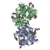
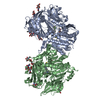
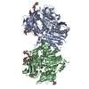
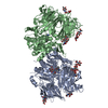
| Deposited unit | 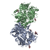
| ||||||||
|---|---|---|---|---|---|---|---|---|---|
| 1 |
| ||||||||
| Unit cell |
|
-Components
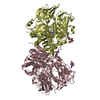
| #1: Protein | Mass: 84462.617 Da / Num. of mol.: 2 Source method: isolated from a genetically manipulated source Source: (gene. exp.) Homo sapiens (human) / Plasmid: pcHDP1-23 / Production host:  Pichia pastoris (fungus) / References: UniProt: P27487, dipeptidyl-peptidase IV Pichia pastoris (fungus) / References: UniProt: P27487, dipeptidyl-peptidase IV#2: Protein/peptide | | Mass: 341.446 Da / Num. of mol.: 1 / Source method: obtained synthetically / Details: This sequence occurs peptide synthesis / Source: (synth.) synthetic construct (others) #3: Sugar | ChemComp-NAG / N-Acetylglucosamine  Type: D-saccharide, beta linking / Mass: 221.208 Da / Num. of mol.: 8 Type: D-saccharide, beta linking / Mass: 221.208 Da / Num. of mol.: 8Source method: isolated from a genetically manipulated source Formula: C8H15NO6 #4: Water | ChemComp-HOH / | Water Mass: 18.015 Da / Num. of mol.: 273 / Source method: isolated from a natural source / Formula: H2O Mass: 18.015 Da / Num. of mol.: 273 / Source method: isolated from a natural source / Formula: H2O |
|---|
-Experimental details
-Experiment
| Experiment | Method: X-RAY DIFFRACTION / Number of used crystals: 1 |
|---|
- Sample preparation
| Crystal | Density Matthews: 2.71 Å3/Da / Density % sol: 54.56 % | ||||||||||||||||||||||||||||||||||||||||||
|---|---|---|---|---|---|---|---|---|---|---|---|---|---|---|---|---|---|---|---|---|---|---|---|---|---|---|---|---|---|---|---|---|---|---|---|---|---|---|---|---|---|---|---|
| Crystal grow | Temperature: 293 K / Method: vapor diffusion, hanging drop / pH: 8.5 Details: Peg, pH 8.5, VAPOR DIFFUSION, HANGING DROP, temperature 293K | ||||||||||||||||||||||||||||||||||||||||||
| Crystal grow | *PLUS Method: vapor diffusion, hanging drop | ||||||||||||||||||||||||||||||||||||||||||
| Components of the solutions | *PLUS
|
-Data collection
| Diffraction | Mean temperature: 90 K |
|---|---|
| Diffraction source | Source: SYNCHROTRON / Site: SLS  / Beamline: X06SA / Wavelength: 0.92 Å / Beamline: X06SA / Wavelength: 0.92 Å |
| Detector | Type: MARRESEARCH / Detector: IMAGE PLATE / Date: Mar 12, 2002 / Details: mirrors |
| Radiation | Monochromator: mirror / Protocol: SINGLE WAVELENGTH / Monochromatic (M) / Laue (L): M / Scattering type: x-ray |
| Radiation wavelength | Wavelength: 0.92 Å / Relative weight: 1 |
| Reflection | Resolution: 2.5→12 Å / Num. all: 171090 / Num. obs: 64208 / % possible obs: 97.5 % / Observed criterion σ(F): 0 / Observed criterion σ(I): 0 |
| Reflection shell | Resolution: 2.5→2.6 Å / % possible all: 99.4 |
| Reflection | *PLUS Num. measured all: 171090 / Rmerge(I) obs: 0.091 |
| Reflection shell | *PLUS % possible obs: 99.4 % / Rmerge(I) obs: 0.159 |
- Processing
| Software |
| ||||||||||||||||||
|---|---|---|---|---|---|---|---|---|---|---|---|---|---|---|---|---|---|---|---|
| Refinement | Method to determine structure: FOURIER SYNTHESIS / Resolution: 2.5→12 Å / σ(F): 0
| ||||||||||||||||||
| Refinement step | Cycle: LAST / Resolution: 2.5→12 Å
| ||||||||||||||||||
| Refinement | *PLUS Lowest resolution: 30 Å | ||||||||||||||||||
| Solvent computation | *PLUS | ||||||||||||||||||
| Displacement parameters | *PLUS | ||||||||||||||||||
| Refine LS restraints | *PLUS
|
