 Movie
Movie Controller


+ Open data

- Basic information
| Entry | Database: PDB / ID: 3zv8 | ||||||
|---|---|---|---|---|---|---|---|
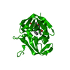
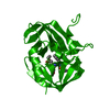
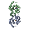
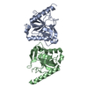
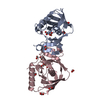
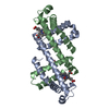
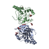
| Title | Crystal structure of 3C protease of Enterovirus 68 | ||||||
 Components Components | 3C PROTEASE Picornain 3C Picornain 3C | ||||||
 Keywords Keywords | HYDROLASE / MICHAEL INHIBITOR | ||||||
| Function / homology |  Function and homology information Function and homology information: / picornain 2A / symbiont-mediated suppression of host mRNA export from nucleus / symbiont genome entry into host cell via pore formation in plasma membrane / picornain 3C / T=pseudo3 icosahedral viral capsid / host cell cytoplasmic vesicle membrane / cytoplasmic vesicle membrane / endocytosis involved in viral entry into host cell / : ...: / picornain 2A / symbiont-mediated suppression of host mRNA export from nucleus / symbiont genome entry into host cell via pore formation in plasma membrane / picornain 3C / T=pseudo3 icosahedral viral capsid / host cell cytoplasmic vesicle membrane / cytoplasmic vesicle membrane / endocytosis involved in viral entry into host cell / : / nucleoside-triphosphate phosphatase / protein complex oligomerization / monoatomic ion channel activity / RNA helicase activity / induction by virus of host autophagy / RNA-directed RNA polymerase / symbiont-mediated suppression of host gene expression / viral RNA genome replication / cysteine-type endopeptidase activity / RNA-dependent RNA polymerase activity / DNA-templated transcription / virion attachment to host cell / structural molecule activity / ATP hydrolysis activity / proteolysis / RNA binding / ATP binding / metal ion bindingSimilarity search - Function | ||||||
| Biological species |  HUMAN ENTEROVIRUS HUMAN ENTEROVIRUS | ||||||
| Method | X-RAY DIFFRACTION / SYNCHROTRON / MOLECULAR REPLACEMENT / Resolution: 2.4 Å | ||||||
 Authors Authors | Tan, J. / Perbandt, M. / Mesters, J.R. / Hilgenfeld, R. | ||||||
 Citation Citation | Journal: J.Virol. / Year: 2013 Title: 3C Protease of Enterovirus 68: Structure-Based Design of Michael Acceptor Inhibitors and Their Broad-Spectrum Antiviral Effects Against Picornaviruses. Authors: Tan, J. / George, S. / Kusov, Y. / Perbandt, M. / Anemuller, S. / Mesters, J.R. / Norder, H. / Coutard, B. / Lacroix, C. / Leyssen, P. / Neyts, J. / Hilgenfeld, R. | ||||||
| History |
| ||||||
| Remark 700 | SHEET DETERMINATION METHOD: DSSP THE SHEETS PRESENTED AS "AA" IN EACH CHAIN ON SHEET RECORDS BELOW ... SHEET DETERMINATION METHOD: DSSP THE SHEETS PRESENTED AS "AA" IN EACH CHAIN ON SHEET RECORDS BELOW IS ACTUALLY AN 6-STRANDED BARREL THIS IS REPRESENTED BY A 7-STRANDED SHEET IN WHICH THE FIRST AND LAST STRANDS ARE IDENTICAL. THE SHEETS PRESENTED AS "AB" IN EACH CHAIN ON SHEET RECORDS BELOW IS ACTUALLY AN 6-STRANDED BARREL THIS IS REPRESENTED BY A 7-STRANDED SHEET IN WHICH THE FIRST AND LAST STRANDS ARE IDENTICAL. |
- Structure visualization
| Structure viewer | Molecule: MolmilJmol/JSmol |
|---|
- Downloads & links
-Download
| PDBx/mmCIF format | 3zv8.cif.gz | 51.2 KB | Display | PDBx/mmCIF format |
|---|---|---|---|---|
| PDB format | pdb3zv8.ent.gz | 36.2 KB | Display | PDB format |
| PDBx/mmJSON format | 3zv8.json.gz | Tree view | PDBx/mmJSON format | |
| Others |  Other downloads Other downloads |
-Validation report
| Arichive directory | https://data.pdbj.org/pub/pdb/validation_reports/zv/3zv8ftp://data.pdbj.org/pub/pdb/validation_reports/zv/3zv8 | HTTPS FTP |
|---|
-Related structure data
| Related structure data | 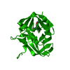 3zv9C  3zvaC  3zvbC 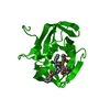 3zvcC  3zvdC  3zveC  3zvfC  3zvgC  1cqqS C: citing same article ( S: Starting model for refinement |
|---|---|
| Similar structure data |


-Links
 PDBj
PDBj

- Assembly
| Deposited unit | 
| ||||||||
|---|---|---|---|---|---|---|---|---|---|
| 1 | 
| ||||||||
| Unit cell |
|
-Components
| #1: Protein | Picornain 3C Mass: 21163.020 Da / Num. of mol.: 1 / Fragment: RESIDUES 1549-1731 Source method: isolated from a genetically manipulated source Source: (gene. exp.) HUMAN ENTEROVIRUS / Strain: 68 / Plasmid: POPINE-EV68 / Production host:  ESCHERICHIA COLI (E. coli) / Strain (production host): BL21 / Variant (production host): TUNER(DE3) PLACI / References: UniProt: A1E4A3, picornain 3C ESCHERICHIA COLI (E. coli) / Strain (production host): BL21 / Variant (production host): TUNER(DE3) PLACI / References: UniProt: A1E4A3, picornain 3C |
|---|---|
| #2: Water | ChemComp-HOH / Water Mass: 18.015 Da / Num. of mol.: 54 / Source method: isolated from a natural source / Formula: H2O Mass: 18.015 Da / Num. of mol.: 54 / Source method: isolated from a natural source / Formula: H2O |
-Experimental details
-Experiment
| Experiment | Method: X-RAY DIFFRACTION / Number of used crystals: 1 |
|---|
- Sample preparation
| Crystal | Density Matthews: 3.73 Å3/Da / Density % sol: 67.05 % / Description: NONE |
|---|---|
| Crystal grow | Method: vapor diffusion, sitting drop / pH: 4.6 Details: 0.07 M SODIUM ACETATE (PH 4.6), 15% PEG 4000, 30% GLYCEROL. SITTING DROP. |
-Data collection
| Diffraction | Mean temperature: 100 K |
|---|---|
| Diffraction source | Source: SYNCHROTRON / Site: BESSY  / Beamline: 14.1 / Wavelength: 0.9184 / Beamline: 14.1 / Wavelength: 0.9184 |
| Detector | Type: MARMOSAIC 225 mm CCD / Detector: CCD / Details: MIRRORS |
| Radiation | Protocol: SINGLE WAVELENGTH / Monochromatic (M) / Laue (L): M / Scattering type: x-ray |
| Radiation wavelength | Wavelength: 0.9184 Å / Relative weight: 1 |
| Reflection | Resolution: 2.4→42.75 Å / Num. obs: 13045 / % possible obs: 100 % / Observed criterion σ(I): 2 / Redundancy: 7.7 % / Rmerge(I) obs: 0.1 / Net I/σ(I): 14.1 |
| Reflection shell | Resolution: 2.4→2.53 Å / Redundancy: 7.9 % / Rmerge(I) obs: 0.57 / Mean I/σ(I) obs: 3.9 / % possible all: 100 |
- Processing
| Software |
| ||||||||||||||||||||||||||||||||||||||||||||||||||||||||||||||||||||||||||||||||||||||||||||||||||||||||||||||||||||||||||||||||||||||||||||||||||||||||||||||||||||||||||||||||||||||
|---|---|---|---|---|---|---|---|---|---|---|---|---|---|---|---|---|---|---|---|---|---|---|---|---|---|---|---|---|---|---|---|---|---|---|---|---|---|---|---|---|---|---|---|---|---|---|---|---|---|---|---|---|---|---|---|---|---|---|---|---|---|---|---|---|---|---|---|---|---|---|---|---|---|---|---|---|---|---|---|---|---|---|---|---|---|---|---|---|---|---|---|---|---|---|---|---|---|---|---|---|---|---|---|---|---|---|---|---|---|---|---|---|---|---|---|---|---|---|---|---|---|---|---|---|---|---|---|---|---|---|---|---|---|---|---|---|---|---|---|---|---|---|---|---|---|---|---|---|---|---|---|---|---|---|---|---|---|---|---|---|---|---|---|---|---|---|---|---|---|---|---|---|---|---|---|---|---|---|---|---|---|---|---|
| Refinement | Method to determine structure: MOLECULAR REPLACEMENT Starting model: PDB ENTRY 1CQQ Resolution: 2.4→42.38 Å / Cor.coef. Fo:Fc: 0.938 / Cor.coef. Fo:Fc free: 0.927 / SU B: 6.15 / SU ML: 0.143 / Cross valid method: THROUGHOUT / ESU R: 0.25 / ESU R Free: 0.204 / Stereochemistry target values: MAXIMUM LIKELIHOOD Details: HYDROGENS HAVE BEEN ADDED IN THE RIDING POSITIONS. U VALUES REFINED INDIVIDUALLY.
| ||||||||||||||||||||||||||||||||||||||||||||||||||||||||||||||||||||||||||||||||||||||||||||||||||||||||||||||||||||||||||||||||||||||||||||||||||||||||||||||||||||||||||||||||||||||
| Solvent computation | Ion probe radii: 0.8 Å / Shrinkage radii: 0.8 Å / VDW probe radii: 1.4 Å / Solvent model: MASK | ||||||||||||||||||||||||||||||||||||||||||||||||||||||||||||||||||||||||||||||||||||||||||||||||||||||||||||||||||||||||||||||||||||||||||||||||||||||||||||||||||||||||||||||||||||||
| Displacement parameters | Biso mean: 35.618 Å2
| ||||||||||||||||||||||||||||||||||||||||||||||||||||||||||||||||||||||||||||||||||||||||||||||||||||||||||||||||||||||||||||||||||||||||||||||||||||||||||||||||||||||||||||||||||||||
| Refinement step | Cycle: LAST / Resolution: 2.4→42.38 Å
| ||||||||||||||||||||||||||||||||||||||||||||||||||||||||||||||||||||||||||||||||||||||||||||||||||||||||||||||||||||||||||||||||||||||||||||||||||||||||||||||||||||||||||||||||||||||
| Refine LS restraints |
|