 Movie
Movie Controller

[English] 日本語
 Yorodumi
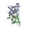
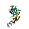
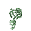
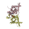
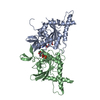
- PDB-3zrh: Crystal structure of the Lys29, Lys33-linkage-specific TRABID OTU... -
+ Open data

- Basic information
| Entry | Database: PDB / ID: 3zrh | ||||||
|---|---|---|---|---|---|---|---|
| Title | Crystal structure of the Lys29, Lys33-linkage-specific TRABID OTU deubiquitinase domain reveals an Ankyrin-repeat ubiquitin binding domain (AnkUBD) | ||||||
 Components Components | UBIQUITIN THIOESTERASE ZRANB1 | ||||||
 Keywords Keywords |  HYDROLASE / DEUBIQUITINATING ENZYME / WNT SIGNALING / OVARIAN TUMOR DOMAIN HYDROLASE / DEUBIQUITINATING ENZYME / WNT SIGNALING / OVARIAN TUMOR DOMAIN | ||||||
| Function / homology |  Function and homology information Function and homology informationprotein K33-linked deubiquitination / protein K29-linked deubiquitination / protein deubiquitination involved in ubiquitin-dependent protein catabolic process / deubiquitinase activity / protein K63-linked deubiquitination / regulation of cell morphogenesis / K63-linked polyubiquitin modification-dependent protein binding / positive regulation of Wnt signaling pathway / polyubiquitin modification-dependent protein binding / cytoskeleton organization ...protein K33-linked deubiquitination / protein K29-linked deubiquitination / protein deubiquitination involved in ubiquitin-dependent protein catabolic process / deubiquitinase activity / protein K63-linked deubiquitination / regulation of cell morphogenesis / K63-linked polyubiquitin modification-dependent protein binding / positive regulation of Wnt signaling pathway / polyubiquitin modification-dependent protein binding / cytoskeleton organization / Degradation of beta-catenin by the destruction complex / Wnt signaling pathway / Ovarian tumor domain proteases / cell migration / ubiquitinyl hydrolase 1 / cysteine-type deubiquitinase activity / intracellular membrane-bounded organelle / nucleoplasm / metal ion binding / nucleus / cytosol / cytoplasmSimilarity search - Function | ||||||
| Biological species |  HOMO SAPIENS (human) HOMO SAPIENS (human) | ||||||
| Method | X-RAY DIFFRACTION / SYNCHROTRON / SIRAS / Resolution: 2.23 Å | ||||||
 Authors Authors | Licchesi, J.D.F. / Akutsu, M. / Komander, D. | ||||||
 Citation Citation | Journal: Nat.Struct.Mol.Biol. / Year: 2011 Title: An Ankyrin-Repeat Ubiquitin-Binding Domain Determines Trabid'S Specificity for Atypical Ubiquitin Chains. Authors: Licchesi, J.D.F. / Mieszczanek, J. / Mevissen, T.E.T. / Rutherford, T.J. / Akutsu, M. / Virdee, S. / Oualid, F.E. / Chin, J.W. / Ovaa, H. / Bienz, M. / Komander, D. | ||||||
| History |
|
- Structure visualization
| Structure viewer | Molecule: MolmilJmol/JSmol |
|---|
- Downloads & links
-Download
| PDBx/mmCIF format | 3zrh.cif.gz | 187.7 KB | Display | PDBx/mmCIF format |
|---|---|---|---|---|
| PDB format | pdb3zrh.ent.gz | 150.3 KB | Display | PDB format |
| PDBx/mmJSON format | 3zrh.json.gz | Tree view | PDBx/mmJSON format | |
| Others |  Other downloads Other downloads |
-Validation report
| Arichive directory | https://data.pdbj.org/pub/pdb/validation_reports/zr/3zrhftp://data.pdbj.org/pub/pdb/validation_reports/zr/3zrh | HTTPS FTP |
|---|
-Related structure data
| Similar structure data |
|---|


-Links
 PDBj
PDBj

- Assembly
| Deposited unit | 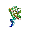
| ||||||||
|---|---|---|---|---|---|---|---|---|---|
| 1 |
| ||||||||
| Unit cell |
|
-Components
| #1: Protein | Mass: 52384.559 Da / Num. of mol.: 1 / Fragment: ANKUBD, OTU, RESIUDES 245-697 Source method: isolated from a genetically manipulated source Source: (gene. exp.) HOMO SAPIENS (human) / Description: ARCTICEXPRESS COMPETENT CELLS / Production host:  ESCHERICHIA COLI (E. coli) / Strain (production host): BL21 / References: UniProt: Q9UGI0, ubiquitinyl hydrolase 1 ESCHERICHIA COLI (E. coli) / Strain (production host): BL21 / References: UniProt: Q9UGI0, ubiquitinyl hydrolase 1 | ||||
|---|---|---|---|---|---|
| #2: Chemical | Ethylene glycol  Mass: 62.068 Da / Num. of mol.: 2 / Source method: obtained synthetically / Formula: C2H6O2 Mass: 62.068 Da / Num. of mol.: 2 / Source method: obtained synthetically / Formula: C2H6O2#3: Chemical | ChemComp-CL / | Chloride  Mass: 35.453 Da / Num. of mol.: 1 / Source method: obtained synthetically / Formula: Cl Mass: 35.453 Da / Num. of mol.: 1 / Source method: obtained synthetically / Formula: Cl#4: Water | ChemComp-HOH / | Water Mass: 18.015 Da / Num. of mol.: 188 / Source method: isolated from a natural source / Formula: H2O Mass: 18.015 Da / Num. of mol.: 188 / Source method: isolated from a natural source / Formula: H2O |
-Experimental details
-Experiment
| Experiment | Method: X-RAY DIFFRACTION / Number of used crystals: 1 |
|---|
- Sample preparation
| Crystal | Density Matthews: 2.77 Å3/Da / Density % sol: 55 % / Description: NONE |
|---|---|
| Crystal grow | pH: 5.9 Details: 150 MM NACL, 100 MM NAOAC, 5 MM MGCL2, 50 MM MES [PH 5.9] |
-Data collection
| Diffraction | Mean temperature: 100 K |
|---|---|
| Diffraction source | Source: SYNCHROTRON / Site: ESRF  / Beamline: ID23-2 / Wavelength: 0.8726 / Beamline: ID23-2 / Wavelength: 0.8726 |
| Detector | Detector: CCD / Date: Nov 29, 2008 |
| Radiation | Protocol: SINGLE WAVELENGTH / Monochromatic (M) / Laue (L): M / Scattering type: x-ray |
| Radiation wavelength | Wavelength: 0.8726 Å / Relative weight: 1 |
| Reflection | Resolution: 2.23→46.32 Å / Num. obs: 29095 / % possible obs: 100 % / Observed criterion σ(I): 2 / Redundancy: 4.1 % / Biso Wilson estimate: 34.45 Å2 / Rmerge(I) obs: 0.08 / Net I/σ(I): 12.1 |
| Reflection shell | Resolution: 2.23→2.35 Å / Redundancy: 4.1 % / Rmerge(I) obs: 0.49 / Mean I/σ(I) obs: 2.7 / % possible all: 100 |
- Processing
| Software |
| |||||||||||||||||||||||||||||||||||||||||||||||||||||||||||||||||||||||||||||
|---|---|---|---|---|---|---|---|---|---|---|---|---|---|---|---|---|---|---|---|---|---|---|---|---|---|---|---|---|---|---|---|---|---|---|---|---|---|---|---|---|---|---|---|---|---|---|---|---|---|---|---|---|---|---|---|---|---|---|---|---|---|---|---|---|---|---|---|---|---|---|---|---|---|---|---|---|---|---|
| Refinement | Method to determine structure: SIRAS Starting model: NONE Resolution: 2.23→44.712 Å / SU ML: 0.85 / σ(F): 0.8 / Phase error: 24.59 / Stereochemistry target values: ML
| |||||||||||||||||||||||||||||||||||||||||||||||||||||||||||||||||||||||||||||
| Solvent computation | Shrinkage radii: 0.9 Å / VDW probe radii: 1.11 Å / Solvent model: FLAT BULK SOLVENT MODEL / Bsol: 44.619 Å2 / ksol: 0.327 e/Å3 | |||||||||||||||||||||||||||||||||||||||||||||||||||||||||||||||||||||||||||||
| Refinement step | Cycle: LAST / Resolution: 2.23→44.712 Å
| |||||||||||||||||||||||||||||||||||||||||||||||||||||||||||||||||||||||||||||
| Refine LS restraints |
| |||||||||||||||||||||||||||||||||||||||||||||||||||||||||||||||||||||||||||||
| LS refinement shell |
| |||||||||||||||||||||||||||||||||||||||||||||||||||||||||||||||||||||||||||||
| Refinement TLS params. | Method: refined / Refine-ID: X-RAY DIFFRACTION
| |||||||||||||||||||||||||||||||||||||||||||||||||||||||||||||||||||||||||||||
| Refinement TLS group |
|
