[English] 日本語
Yorodumi
- PDB-3zpq: Thermostabilised turkey beta1 adrenergic receptor with 4-(piperaz... -

+
Open data


ID or keywords:

Loading...

-
Basic information

Entry
Database: PDB / ID: 3zpq
TitleThermostabilised turkey beta1 adrenergic receptor with 4-(piperazin-1- yl)-1H-indole bound (compound 19)
ComponentsBETA-1 ADRENERGIC RECEPTOR
KeywordsMEMBRANE PROTEIN / GPCR / FRAGMENT SCREENING / ARYL PIPERAZINE / STRUCTURE BASED DRUG DESIGN
Function / homology
Function and homology information


beta1-adrenergic receptor activity / positive regulation of heart contraction / regulation of circadian sleep/wake cycle, sleep / adenylate cyclase-activating adrenergic receptor signaling pathway / positive regulation of GTPase activity / early endosome / membrane / identical protein binding / plasma membrane
Similarity search - Function
Beta 1 adrenoceptor / Adrenoceptor family / Rhopdopsin 7-helix transmembrane proteins / Rhodopsin 7-helix transmembrane proteins / Serpentine type 7TM GPCR chemoreceptor Srsx / G-protein coupled receptors family 1 signature. / G protein-coupled receptor, rhodopsin-like / GPCR, rhodopsin-like, 7TM / G-protein coupled receptors family 1 profile. / 7 transmembrane receptor (rhodopsin family) ...Beta 1 adrenoceptor / Adrenoceptor family / Rhopdopsin 7-helix transmembrane proteins / Rhodopsin 7-helix transmembrane proteins / Serpentine type 7TM GPCR chemoreceptor Srsx / G-protein coupled receptors family 1 signature. / G protein-coupled receptor, rhodopsin-like / GPCR, rhodopsin-like, 7TM / G-protein coupled receptors family 1 profile. / 7 transmembrane receptor (rhodopsin family) / Up-down Bundle / Mainly Alpha
Similarity search - Domain/homology
4-(PIPERAZIN-1-YL)-1H-INDOLE / CHOLESTEROL HEMISUCCINATE / Beta-1 adrenergic receptor
Similarity search - Component
Biological speciesMELEAGRIS GALLOPAVO (turkey)
MethodX-RAY DIFFRACTION / SYNCHROTRON / MOLECULAR REPLACEMENT / Resolution: 2.8 Å
AuthorsChristopher, J.A. / Congreve, M. / Dore, A.S. / Marshall, F.H. / Myszka, D.G. / Brown, J. / Koglin, M. / Tehan, B. / Errey, J.C. / Tate, C.G. / Warne, T.
CitationJournal: J.Med.Chem. / Year: 2013
Title: Biophysical Fragment Screening of the Beta1-Adrenergic Receptor: Identification of High Affinity Aryl Piperazine Leads Using Structure-Based Drug Design.
Authors: Christopher, J. / Brown, J. / Dore, A. / Errey, J. / Koglin, M. / Marshall, F.H. / Myszka, D. / Rich, R.L. / Tate, C.G. / Tehan, B. / Warne, T. / Congreve, M.
History
DepositionMar 1, 2013Deposition site: PDBE / Processing site: PDBE
Revision 1.0Apr 3, 2013Provider: repository / Type: Initial release
Revision 1.1May 22, 2013Group: Database references
Revision 1.2Apr 3, 2019Group: Data collection / Other / Source and taxonomy
Category: entity_src_gen / pdbx_database_proc / pdbx_database_status
Item: _entity_src_gen.pdbx_host_org_cell_line / _pdbx_database_status.recvd_author_approval
Revision 1.3May 15, 2019Group: Data collection / Experimental preparation / Category: exptl_crystal_grow / Item: _exptl_crystal_grow.temp
Revision 1.4Dec 20, 2023Group: Data collection / Database references ...Data collection / Database references / Derived calculations / Other / Refinement description
Category: chem_comp_atom / chem_comp_bond ...chem_comp_atom / chem_comp_bond / database_2 / pdbx_database_status / pdbx_initial_refinement_model / pdbx_struct_conn_angle / struct_conn / struct_site
Item: _database_2.pdbx_DOI / _database_2.pdbx_database_accession ..._database_2.pdbx_DOI / _database_2.pdbx_database_accession / _pdbx_database_status.status_code_sf / _pdbx_struct_conn_angle.ptnr1_auth_comp_id / _pdbx_struct_conn_angle.ptnr1_auth_seq_id / _pdbx_struct_conn_angle.ptnr1_label_asym_id / _pdbx_struct_conn_angle.ptnr1_label_atom_id / _pdbx_struct_conn_angle.ptnr1_label_comp_id / _pdbx_struct_conn_angle.ptnr1_label_seq_id / _pdbx_struct_conn_angle.ptnr3_auth_comp_id / _pdbx_struct_conn_angle.ptnr3_auth_seq_id / _pdbx_struct_conn_angle.ptnr3_label_asym_id / _pdbx_struct_conn_angle.ptnr3_label_atom_id / _pdbx_struct_conn_angle.ptnr3_label_comp_id / _pdbx_struct_conn_angle.ptnr3_label_seq_id / _pdbx_struct_conn_angle.value / _struct_conn.pdbx_dist_value / _struct_conn.ptnr1_auth_comp_id / _struct_conn.ptnr1_auth_seq_id / _struct_conn.ptnr1_label_asym_id / _struct_conn.ptnr1_label_atom_id / _struct_conn.ptnr1_label_comp_id / _struct_conn.ptnr1_label_seq_id / _struct_conn.ptnr2_auth_comp_id / _struct_conn.ptnr2_auth_seq_id / _struct_conn.ptnr2_label_asym_id / _struct_conn.ptnr2_label_atom_id / _struct_conn.ptnr2_label_comp_id / _struct_conn.ptnr2_label_seq_id / _struct_site.pdbx_auth_asym_id / _struct_site.pdbx_auth_comp_id / _struct_site.pdbx_auth_seq_id

-
Structure visualization

Structure viewerMolecule:
MolmilJmol/JSmol

Downloads & links

-
Assembly

Deposited unit
A: BETA-1 ADRENERGIC RECEPTOR
B: BETA-1 ADRENERGIC RECEPTOR
hetero molecules


Theoretical massNumber of molelcules
Total (without water)77,71520
Polymers71,8822
Non-polymers5,83418
Water63135
1
A: BETA-1 ADRENERGIC RECEPTOR
hetero molecules


Theoretical massNumber of molelcules
Total (without water)38,6569
Polymers35,9411
Non-polymers2,7168
Water181
TypeNameSymmetry operationNumber
identity operation1_555x,y,z1
MethodPISA
2
B: BETA-1 ADRENERGIC RECEPTOR
hetero molecules


Theoretical massNumber of molelcules
Total (without water)39,05911
Polymers35,9411
Non-polymers3,11810
Water181
TypeNameSymmetry operationNumber
identity operation1_555x,y,z1
MethodPISA
Unit cell
Length a, b, c (Å)89.852, 61.441, 100.784
Angle α, β, γ (deg.)90.00, 108.89, 90.00
Int Tables number4
Space group name H-MP1211
Noncrystallographic symmetry (NCS)NCS oper:
IDCodeMatrixVector
1given(1), (1), (1)
2given(-0.999991, 0.004062, 0.001058), (0.003701, 0.734531, 0.678565), (0.001979, 0.678563, -0.734539)-13.73911, -18.27991, 46.50096

-
Components

-
Protein , 1 types, 2 molecules AB

#1: Protein BETA-1 ADRENERGIC RECEPTOR / / BETA1 ADRENERGIC RECEPTOR / BETA-1 ADRENORECEPTOR / BETA-1 ADRENOCEPTOR / BETA-T


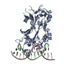
Mass: 35940.777 Da / Num. of mol.: 2 / Fragment: RESIDUES 33-243,272-368 / Mutation: YES
Source method: isolated from a genetically manipulated source
Details: RESIDUES 3-32 AT THE N-TERMINUS AND RESIDUES 244-271 OF THE THIRD INTRACELLULAR LOOP WERE DELETED FROM THE CONSTRUCT. THE CONSTRUCT WAS TRUNCATED AFTER RESIDUE 367 AND A HEXAHIS TAG ADDED.
Source: (gene. exp.) MELEAGRIS GALLOPAVO (turkey) / Cell: ERYTHROCYTE / Plasmid: PBACPAK8 / Cell line (production host): High Five / Production host: TRICHOPLUSIA NI (cabbage looper) / References: UniProt: P07700

-
Non-polymers , 5 types, 53 molecules

#2: Chemical
ChemComp-Y01 / CHOLESTEROL HEMISUCCINATE


Mass: 486.726 Da / Num. of mol.: 4 / Source method: obtained synthetically / Formula: C31H50O4
#3: Chemical
ChemComp-2CV / HEGA-10


Mass: 379.489 Da / Num. of mol.: 9 / Source method: obtained synthetically / Formula: C18H37NO7 / Comment: detergent*YM
#4: Chemical ChemComp-XF5 / 4-(PIPERAZIN-1-YL)-1H-INDOLE


Mass: 201.268 Da / Num. of mol.: 2 / Source method: obtained synthetically / Formula: C12H15N3
#5: Chemical ChemComp-NA / SODIUM ION


Mass: 22.990 Da / Num. of mol.: 3 / Source method: obtained synthetically / Formula: Na
#6: Water ChemComp-HOH / water / Water


Mass: 18.015 Da / Num. of mol.: 35 / Source method: isolated from a natural source / Formula: H2O

-
Details

Sequence detailsTHE FOLLOWING MUTATIONS WERE MADE TO IMPROVE THERMOSTABILITY R68S,M90V,Y227A,A282L,F327A,F338M THE ...THE FOLLOWING MUTATIONS WERE MADE TO IMPROVE THERMOSTABILITY R68S,M90V,Y227A,A282L,F327A,F338M THE FOLLOWING MUTATIONS WERE MADE TO IMPROVE EXPRESSION AND HELP CRYSTALLISATION C116L, C358A

-
Experimental details

-
Experiment

ExperimentMethod: X-RAY DIFFRACTION / Number of used crystals: 1

-
Sample preparation

CrystalDensity Matthews: 3.66 Å3/Da / Density % sol: 66.37 % / Description: NONE
Crystal growTemperature: 277 K / pH: 9 / Details: 0.1M BICINE PH9.0, 24% PEG600, 4C

-
Data collection

DiffractionMean temperature: 100 K
Diffraction sourceSource: SYNCHROTRON / Site: Diamond / Beamline: I24 / Wavelength: 0.9778
DetectorType: DECTRIS PILATUS 6M / Detector: PIXEL / Date: Feb 2, 2012
RadiationProtocol: SINGLE WAVELENGTH / Monochromatic (M) / Laue (L): M / Scattering type: x-ray
Radiation wavelengthWavelength: 0.9778 Å / Relative weight: 1
ReflectionResolution: 2.8→61.4 Å / Num. obs: 23841 / % possible obs: 92.5 % / Observed criterion σ(I): 2 / Redundancy: 3.3 % / Rmerge(I) obs: 0.18 / Net I/σ(I): 6.8
Reflection shellResolution: 2.8→2.95 Å / Redundancy: 2.7 % / Rmerge(I) obs: 0.65 / Mean I/σ(I) obs: 1.9 / % possible all: 90.9

-
Processing

Software
NameVersionClassification
REFMAC5.6.0117refinement
MOSFLMdata reduction
SCALAdata scaling
PHASERphasing
RefinementMethod to determine structure: MOLECULAR REPLACEMENT
Starting model: PDB ENTRY 4AMJ
Resolution: 2.8→95.36 Å / Cor.coef. Fo:Fc: 0.906 / Cor.coef. Fo:Fc free: 0.86 / SU B: 11.771 / SU ML: 0.234 / Cross valid method: THROUGHOUT / σ(F): 2 / ESU R: 0.89 / ESU R Free: 0.377 / Stereochemistry target values: MAXIMUM LIKELIHOOD / Details: HYDROGENS HAVE BEEN ADDED IN THE RIDING POSITIONS.
RfactorNum. reflection% reflection
Rfree0.2737 1214 5.1 %
Rwork0.22026 --
obs0.22298 22615 91.74 %
Solvent computationIon probe radii: 0.8 Å / Shrinkage radii: 0.8 Å / VDW probe radii: 1.2 Å / Solvent model: MASK
Displacement parametersBiso mean: 45.24 Å2
Baniso -1Baniso -2Baniso -3
1-0.06 Å20 Å20.94 Å2
2--1.82 Å20 Å2
3----1.27 Å2
Refinement stepCycle: LAST / Resolution: 2.8→95.36 Å
ProteinNucleic acidLigandSolventTotal
Num. atoms4648 0 313 35 4996
Refine LS restraints
Refine-IDTypeDev idealDev ideal targetNumber
X-RAY DIFFRACTIONr_bond_refined_d0.0090.025090
X-RAY DIFFRACTIONr_bond_other_d0.0030.023533
X-RAY DIFFRACTIONr_angle_refined_deg1.2972.0086903
X-RAY DIFFRACTIONr_angle_other_deg0.9663.0188120
X-RAY DIFFRACTIONr_dihedral_angle_1_deg4.8465583
X-RAY DIFFRACTIONr_dihedral_angle_2_deg34.01722.022178
X-RAY DIFFRACTIONr_dihedral_angle_3_deg15.44315808
X-RAY DIFFRACTIONr_dihedral_angle_4_deg17.3051535
X-RAY DIFFRACTIONr_chiral_restr0.0630.2819
X-RAY DIFFRACTIONr_gen_planes_refined0.0050.0215258
X-RAY DIFFRACTIONr_gen_planes_other0.0020.021067
X-RAY DIFFRACTIONr_nbd_refined
X-RAY DIFFRACTIONr_nbd_other
X-RAY DIFFRACTIONr_nbtor_refined
X-RAY DIFFRACTIONr_nbtor_other
X-RAY DIFFRACTIONr_xyhbond_nbd_refined
X-RAY DIFFRACTIONr_xyhbond_nbd_other
X-RAY DIFFRACTIONr_metal_ion_refined
X-RAY DIFFRACTIONr_metal_ion_other
X-RAY DIFFRACTIONr_symmetry_vdw_refined
X-RAY DIFFRACTIONr_symmetry_vdw_other
X-RAY DIFFRACTIONr_symmetry_hbond_refined
X-RAY DIFFRACTIONr_symmetry_hbond_other
X-RAY DIFFRACTIONr_symmetry_metal_ion_refined
X-RAY DIFFRACTIONr_symmetry_metal_ion_other
X-RAY DIFFRACTIONr_mcbond_it
X-RAY DIFFRACTIONr_mcbond_other
X-RAY DIFFRACTIONr_mcangle_it
X-RAY DIFFRACTIONr_mcangle_other
X-RAY DIFFRACTIONr_scbond_it
X-RAY DIFFRACTIONr_scbond_other
X-RAY DIFFRACTIONr_scangle_it
X-RAY DIFFRACTIONr_scangle_other
X-RAY DIFFRACTIONr_long_range_B_refined
X-RAY DIFFRACTIONr_long_range_B_other
X-RAY DIFFRACTIONr_rigid_bond_restr
X-RAY DIFFRACTIONr_sphericity_free
X-RAY DIFFRACTIONr_sphericity_bonded
LS refinement shellResolution: 2.8→2.873 Å / Total num. of bins used: 20
RfactorNum. reflection% reflection
Rfree0.354 89 -
Rwork0.278 1555 -
obs--90.03 %

+
About Yorodumi

-
News

-
Feb 9, 2022. New format data for meta-information of EMDB entries

New format data for meta-information of EMDB entries

  • Version 3 of the EMDB header file is now the official format.
  • The previous official version 1.9 will be removed from the archive.

Related info.:EMDB header

External links:wwPDB to switch to version 3 of the EMDB data model

-
Aug 12, 2020. Covid-19 info

Covid-19 info

URL: https://pdbj.org/emnavi/covid19.php

New page: Covid-19 featured information page in EM Navigator.

Related info.:Covid-19 info / Mar 5, 2020. Novel coronavirus structure data

+
Mar 5, 2020. Novel coronavirus structure data

Novel coronavirus structure data

Related info.:Yorodumi Speices / Aug 12, 2020. Covid-19 info

External links:COVID-19 featured content - PDBj / Molecule of the Month (242):Coronavirus Proteases

+
Jan 31, 2019. EMDB accession codes are about to change! (news from PDBe EMDB page)

EMDB accession codes are about to change! (news from PDBe EMDB page)

  • The allocation of 4 digits for EMDB accession codes will soon come to an end. Whilst these codes will remain in use, new EMDB accession codes will include an additional digit and will expand incrementally as the available range of codes is exhausted. The current 4-digit format prefixed with “EMD-” (i.e. EMD-XXXX) will advance to a 5-digit format (i.e. EMD-XXXXX), and so on. It is currently estimated that the 4-digit codes will be depleted around Spring 2019, at which point the 5-digit format will come into force.
  • The EM Navigator/Yorodumi systems omit the EMD- prefix.

Related info.:Q: What is EMD? / ID/Accession-code notation in Yorodumi/EM Navigator

External links:EMDB Accession Codes are Changing Soon! / Contact to PDBj

+
Jul 12, 2017. Major update of PDB

Major update of PDB

  • wwPDB released updated PDB data conforming to the new PDBx/mmCIF dictionary.
  • This is a major update changing the version number from 4 to 5, and with Remediation, in which all the entries are updated.
  • In this update, many items about electron microscopy experimental information are reorganized (e.g. em_software).
  • Now, EM Navigator and Yorodumi are based on the updated data.

External links:wwPDB Remediation / Enriched Model Files Conforming to OneDep Data Standards Now Available in the PDB FTP Archive

-
Yorodumi

Thousand views of thousand structures

  • Yorodumi is a browser for structure data from EMDB, PDB, SASBDB, etc.
  • This page is also the successor to EM Navigator detail page, and also detail information page/front-end page for Omokage search.
  • The word "yorodu" (or yorozu) is an old Japanese word meaning "ten thousand". "mi" (miru) is to see.

Related info.:EMDB / PDB / SASBDB / Comparison of 3 databanks / Yorodumi Search / Aug 31, 2016. New EM Navigator & Yorodumi / Yorodumi Papers / Jmol/JSmol / Function and homology information / Changes in new EM Navigator and Yorodumi

Read more