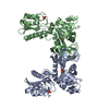
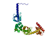
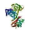
[English] 日本語
Yorodumi
- PDB-3zpg: Acinetobacter baumannii RibD, form 2 -

+
Open data


ID or keywords:

Loading...

-
Basic information

Entry
Database: PDB / ID: 3zpg
TitleAcinetobacter baumannii RibD, form 2
ComponentsRIBD
KeywordsHYDROLASE / REDUCTASE / DEAMINASE
Function / homology
Function and homology information


5-amino-6-(5-phosphoribosylamino)uracil reductase / diaminohydroxyphosphoribosylaminopyrimidine deaminase / diaminohydroxyphosphoribosylaminopyrimidine deaminase activity / 5-amino-6-(5-phosphoribosylamino)uracil reductase activity / riboflavin biosynthetic process / NADP binding / zinc ion binding
Similarity search - Function
Riboflavin biosynthesis protein RibD / Riboflavin-specific deaminase, C-terminal / Bacterial bifunctional deaminase-reductase, C-terminal / RibD C-terminal domain / Cytidine and deoxycytidylate deaminase zinc-binding region / Cytidine Deaminase, domain 2 / Cytidine Deaminase; domain 2 / APOBEC/CMP deaminase, zinc-binding / Cytidine and deoxycytidylate deaminases zinc-binding region signature. / Cytidine and deoxycytidylate deaminase domain ...Riboflavin biosynthesis protein RibD / Riboflavin-specific deaminase, C-terminal / Bacterial bifunctional deaminase-reductase, C-terminal / RibD C-terminal domain / Cytidine and deoxycytidylate deaminase zinc-binding region / Cytidine Deaminase, domain 2 / Cytidine Deaminase; domain 2 / APOBEC/CMP deaminase, zinc-binding / Cytidine and deoxycytidylate deaminases zinc-binding region signature. / Cytidine and deoxycytidylate deaminase domain / Cytidine and deoxycytidylate deaminases domain profile. / Cytidine deaminase-like / Dihydrofolate Reductase, subunit A / Dihydrofolate Reductase, subunit A / Dihydrofolate reductase-like domain superfamily / 3-Layer(aba) Sandwich / Alpha Beta
Similarity search - Domain/homology
GUANOSINE-5'-MONOPHOSPHATE / ACETATE ION / CACODYLATE ION / OXALATE ION / Riboflavin biosynthesis protein RibD
Similarity search - Component
Biological speciesACINETOBACTER BAUMANNII (bacteria)
MethodX-RAY DIFFRACTION / MOLECULAR REPLACEMENT / Resolution: 1.99 Å
AuthorsDawson, A. / Trumper, P. / Chrysostomou, G. / Hunter, W.N.
CitationJournal: Acta Crystallogr.,Sect.F / Year: 2013
Title: Structure of Diaminohydroxyphosphoribosylaminopyrimidine Deaminase/5-Amino-6-(5-Phosphoribosylamino)Uracil Reductase from Acinetobacter Baumannii.
Authors: Dawson, A. / Trumper, P. / Chrysostomou, G. / Hunter, W.N.
History
DepositionFeb 27, 2013Deposition site: PDBE / Processing site: PDBE
Revision 1.0Mar 27, 2013Provider: repository / Type: Initial release
Revision 1.1Jun 12, 2013Group: Database references
Revision 1.2Jun 28, 2017Group: Data collection / Category: diffrn_source / Item: _diffrn_source.type
Revision 1.3Dec 20, 2023Group: Data collection / Database references ...Data collection / Database references / Derived calculations / Other / Refinement description
Category: chem_comp_atom / chem_comp_bond ...chem_comp_atom / chem_comp_bond / database_2 / pdbx_database_status / pdbx_initial_refinement_model / pdbx_struct_conn_angle / struct_conn / struct_site
Item: _database_2.pdbx_DOI / _database_2.pdbx_database_accession ..._database_2.pdbx_DOI / _database_2.pdbx_database_accession / _pdbx_database_status.status_code_sf / _pdbx_struct_conn_angle.ptnr1_auth_seq_id / _pdbx_struct_conn_angle.ptnr1_label_seq_id / _pdbx_struct_conn_angle.ptnr3_auth_seq_id / _pdbx_struct_conn_angle.ptnr3_label_seq_id / _pdbx_struct_conn_angle.value / _struct_conn.pdbx_dist_value / _struct_conn.ptnr1_auth_comp_id / _struct_conn.ptnr1_auth_seq_id / _struct_conn.ptnr1_label_asym_id / _struct_conn.ptnr1_label_atom_id / _struct_conn.ptnr1_label_comp_id / _struct_conn.ptnr1_label_seq_id / _struct_conn.ptnr2_auth_comp_id / _struct_conn.ptnr2_auth_seq_id / _struct_conn.ptnr2_label_asym_id / _struct_conn.ptnr2_label_atom_id / _struct_conn.ptnr2_label_comp_id / _struct_conn.ptnr2_label_seq_id / _struct_site.pdbx_auth_asym_id / _struct_site.pdbx_auth_comp_id / _struct_site.pdbx_auth_seq_id

-
Structure visualization

Structure viewerMolecule:
MolmilJmol/JSmol

Downloads & links

-
Assembly

Deposited unit
A: RIBD
hetero molecules


Theoretical massNumber of molelcules
Total (without water)43,47911
Polymers42,0871
Non-polymers1,39210
Water2,288127
1
A: RIBD
hetero molecules

A: RIBD
hetero molecules


Theoretical massNumber of molelcules
Total (without water)86,95822
Polymers84,1752
Non-polymers2,78320
Water362
TypeNameSymmetry operationNumber
identity operation1_555x,y,z1
crystal symmetry operation7_555y,x,-z1
Buried area9310 Å2
ΔGint-177.5 kcal/mol
Surface area30680 Å2
MethodPISA
Unit cell
Length a, b, c (Å)72.998, 72.998, 201.456
Angle α, β, γ (deg.)90.00, 90.00, 90.00
Int Tables number96
Space group name H-MP43212

-
Components

-
Protein , 1 types, 1 molecules A

#1: Protein RIBD


Mass: 42087.293 Da / Num. of mol.: 1
Source method: isolated from a genetically manipulated source
Source: (gene. exp.) ACINETOBACTER BAUMANNII (bacteria) / Plasmid: PET15BTEV / Production host: ESCHERICHIA COLI (E. coli) / Strain (production host): BL21(DE3)
References: UniProt: D0CB74, diaminohydroxyphosphoribosylaminopyrimidine deaminase, 5-amino-6-(5-phosphoribosylamino)uracil reductase

-
Non-polymers , 8 types, 137 molecules

#2: Chemical ChemComp-ZN / ZINC ION


Mass: 65.409 Da / Num. of mol.: 1 / Source method: obtained synthetically / Formula: Zn
#3: Chemical ChemComp-SO4 / SULFATE ION / Sulfate


Mass: 96.063 Da / Num. of mol.: 2 / Source method: obtained synthetically / Formula: SO4
#4: Chemical ChemComp-5GP / GUANOSINE-5'-MONOPHOSPHATE / Guanosine monophosphate


Mass: 363.221 Da / Num. of mol.: 2 / Source method: obtained synthetically / Formula: C10H14N5O8P
#5: Chemical ChemComp-OXL / OXALATE ION / Oxalate


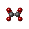
Mass: 88.019 Da / Num. of mol.: 2 / Source method: obtained synthetically / Formula: C2O4
#6: Chemical ChemComp-CAC / CACODYLATE ION / dimethylarsinate / Cacodylic acid


Mass: 136.989 Da / Num. of mol.: 1 / Source method: obtained synthetically / Formula: C2H6AsO2
#7: Chemical ChemComp-CL / CHLORIDE ION / Chloride


Mass: 35.453 Da / Num. of mol.: 1 / Source method: obtained synthetically / Formula: Cl
#8: Chemical ChemComp-ACT / ACETATE ION / Acetate


Mass: 59.044 Da / Num. of mol.: 1 / Source method: obtained synthetically / Formula: C2H3O2
#9: Water ChemComp-HOH / water / Water


Mass: 18.015 Da / Num. of mol.: 127 / Source method: isolated from a natural source / Formula: H2O

-
Experimental details

-
Experiment

ExperimentMethod: X-RAY DIFFRACTION / Number of used crystals: 1

-
Sample preparation

CrystalDensity Matthews: 3.19 Å3/Da / Density % sol: 61.53 % / Description: NONE
Crystal growDetails: 0.2 M AMMONIUM SULFATE, 0.125 M SODIUM CACODYLATE, 30% W/V POLYETHYLENE GLYCOL 8000

-
Data collection

DiffractionMean temperature: 100 K
Diffraction sourceSource: ROTATING ANODE / Type: RIGAKU MICROMAX-007 HF / Wavelength: 1.5418
DetectorType: RIGAKU IMAGE PLATE / Detector: IMAGE PLATE / Date: Aug 15, 2011 / Details: MIRRORS
RadiationMonochromator: SI / Protocol: SINGLE WAVELENGTH / Monochromatic (M) / Laue (L): M / Scattering type: x-ray
Radiation wavelengthWavelength: 1.5418 Å / Relative weight: 1
ReflectionResolution: 1.99→51.62 Å / Num. obs: 38040 / % possible obs: 99.5 % / Observed criterion σ(I): 0 / Redundancy: 13.3 % / Rmerge(I) obs: 0.06 / Net I/σ(I): 24.7
Reflection shellResolution: 1.99→2.1 Å / Redundancy: 12 % / Rmerge(I) obs: 0.62 / Mean I/σ(I) obs: 4.1 / % possible all: 97

-
Processing

Software
NameVersionClassification
REFMAC5.5.0109refinement
XDSdata reduction
SCALAdata scaling
PHASERphasing
RefinementMethod to determine structure: MOLECULAR REPLACEMENT
Starting model: PDB ENTRY 3ZPC
Resolution: 1.99→68.63 Å / Cor.coef. Fo:Fc: 0.947 / Cor.coef. Fo:Fc free: 0.941 / SU B: 7.342 / SU ML: 0.106 / Cross valid method: THROUGHOUT / ESU R: 0.152 / ESU R Free: 0.138 / Stereochemistry target values: MAXIMUM LIKELIHOOD / Details: HYDROGENS HAVE BEEN ADDED IN THE RIDING POSITIONS.
RfactorNum. reflection% reflectionSelection details
Rfree0.23671 1897 5 %RANDOM
Rwork0.21393 ---
obs0.21508 36017 99.4 %-
Solvent computationIon probe radii: 0.8 Å / Shrinkage radii: 0.8 Å / VDW probe radii: 1.4 Å / Solvent model: MASK
Displacement parametersBiso mean: 44.915 Å2
Baniso -1Baniso -2Baniso -3
1-0.52 Å20 Å20 Å2
2--0.52 Å20 Å2
3----1.05 Å2
Refinement stepCycle: LAST / Resolution: 1.99→68.63 Å
ProteinNucleic acidLigandSolventTotal
Num. atoms2714 0 81 127 2922
Refine LS restraints
Refine-IDTypeDev idealDev ideal targetNumber
X-RAY DIFFRACTIONr_bond_refined_d0.010.0222874
X-RAY DIFFRACTIONr_bond_other_d
X-RAY DIFFRACTIONr_angle_refined_deg1.2882.0113908
X-RAY DIFFRACTIONr_angle_other_deg
X-RAY DIFFRACTIONr_dihedral_angle_1_deg5.6315356
X-RAY DIFFRACTIONr_dihedral_angle_2_deg43.65224.918122
X-RAY DIFFRACTIONr_dihedral_angle_3_deg14.41115507
X-RAY DIFFRACTIONr_dihedral_angle_4_deg20.4531519
X-RAY DIFFRACTIONr_chiral_restr0.0770.2455
X-RAY DIFFRACTIONr_gen_planes_refined0.0050.0212122
X-RAY DIFFRACTIONr_gen_planes_other
X-RAY DIFFRACTIONr_nbd_refined
X-RAY DIFFRACTIONr_nbd_other
X-RAY DIFFRACTIONr_nbtor_refined
X-RAY DIFFRACTIONr_nbtor_other
X-RAY DIFFRACTIONr_xyhbond_nbd_refined
X-RAY DIFFRACTIONr_xyhbond_nbd_other
X-RAY DIFFRACTIONr_metal_ion_refined
X-RAY DIFFRACTIONr_metal_ion_other
X-RAY DIFFRACTIONr_symmetry_vdw_refined
X-RAY DIFFRACTIONr_symmetry_vdw_other
X-RAY DIFFRACTIONr_symmetry_hbond_refined
X-RAY DIFFRACTIONr_symmetry_hbond_other
X-RAY DIFFRACTIONr_symmetry_metal_ion_refined
X-RAY DIFFRACTIONr_symmetry_metal_ion_other
X-RAY DIFFRACTIONr_mcbond_it0.5971.51775
X-RAY DIFFRACTIONr_mcbond_other
X-RAY DIFFRACTIONr_mcangle_it1.10922865
X-RAY DIFFRACTIONr_mcangle_other
X-RAY DIFFRACTIONr_scbond_it1.7731099
X-RAY DIFFRACTIONr_scbond_other
X-RAY DIFFRACTIONr_scangle_it2.8084.51041
X-RAY DIFFRACTIONr_scangle_other
X-RAY DIFFRACTIONr_long_range_B_refined
X-RAY DIFFRACTIONr_long_range_B_other
X-RAY DIFFRACTIONr_rigid_bond_restr
X-RAY DIFFRACTIONr_sphericity_free
X-RAY DIFFRACTIONr_sphericity_bonded
LS refinement shellResolution: 1.994→2.046 Å / Total num. of bins used: 20
RfactorNum. reflection% reflection
Rfree0.324 128 -
Rwork0.294 2455 -
obs--93.82 %
Refinement TLS params.

Method: refined / Refine-ID: X-RAY DIFFRACTION

IDL112)L122)L132)L222)L232)L332)S11 (Å °)S12 (Å °)S13 (Å °)S21 (Å °)S22 (Å °)S23 (Å °)S31 (Å °)S32 (Å °)S33 (Å °)T112)T122)T132)T222)T232)T332)Origin x (Å)Origin y (Å)Origin z (Å)
16.00011.8534-1.37064.3068-1.10841.3729-0.0627-0.129-0.0524-0.4601-0.0525-0.9207-0.04910.50830.11520.3186-0.07780.30810.2526-0.00640.536413.595-14.12-24.565
20.9920.0868-0.03681.68060.45251.3184-0.1354-0.0137-0.1464-0.40350.012-0.23120.04580.08950.12340.26470.02710.11730.030.0140.2273-9.776-25.72-17.707
Refinement TLS group
IDRefine-IDRefine TLS-IDAuth asym-IDAuth seq-ID
1X-RAY DIFFRACTION1A2 - 88
2X-RAY DIFFRACTION2A89 - 357

+
About Yorodumi

-
News

-
Feb 9, 2022. New format data for meta-information of EMDB entries

New format data for meta-information of EMDB entries

  • Version 3 of the EMDB header file is now the official format.
  • The previous official version 1.9 will be removed from the archive.

Related info.:EMDB header

External links:wwPDB to switch to version 3 of the EMDB data model

-
Aug 12, 2020. Covid-19 info

Covid-19 info

URL: https://pdbj.org/emnavi/covid19.php

New page: Covid-19 featured information page in EM Navigator.

Related info.:Covid-19 info / Mar 5, 2020. Novel coronavirus structure data

+
Mar 5, 2020. Novel coronavirus structure data

Novel coronavirus structure data

Related info.:Yorodumi Speices / Aug 12, 2020. Covid-19 info

External links:COVID-19 featured content - PDBj / Molecule of the Month (242):Coronavirus Proteases

+
Jan 31, 2019. EMDB accession codes are about to change! (news from PDBe EMDB page)

EMDB accession codes are about to change! (news from PDBe EMDB page)

  • The allocation of 4 digits for EMDB accession codes will soon come to an end. Whilst these codes will remain in use, new EMDB accession codes will include an additional digit and will expand incrementally as the available range of codes is exhausted. The current 4-digit format prefixed with “EMD-” (i.e. EMD-XXXX) will advance to a 5-digit format (i.e. EMD-XXXXX), and so on. It is currently estimated that the 4-digit codes will be depleted around Spring 2019, at which point the 5-digit format will come into force.
  • The EM Navigator/Yorodumi systems omit the EMD- prefix.

Related info.:Q: What is EMD? / ID/Accession-code notation in Yorodumi/EM Navigator

External links:EMDB Accession Codes are Changing Soon! / Contact to PDBj

+
Jul 12, 2017. Major update of PDB

Major update of PDB

  • wwPDB released updated PDB data conforming to the new PDBx/mmCIF dictionary.
  • This is a major update changing the version number from 4 to 5, and with Remediation, in which all the entries are updated.
  • In this update, many items about electron microscopy experimental information are reorganized (e.g. em_software).
  • Now, EM Navigator and Yorodumi are based on the updated data.

External links:wwPDB Remediation / Enriched Model Files Conforming to OneDep Data Standards Now Available in the PDB FTP Archive

-
Yorodumi

Thousand views of thousand structures

  • Yorodumi is a browser for structure data from EMDB, PDB, SASBDB, etc.
  • This page is also the successor to EM Navigator detail page, and also detail information page/front-end page for Omokage search.
  • The word "yorodu" (or yorozu) is an old Japanese word meaning "ten thousand". "mi" (miru) is to see.

Related info.:EMDB / PDB / SASBDB / Comparison of 3 databanks / Yorodumi Search / Aug 31, 2016. New EM Navigator & Yorodumi / Yorodumi Papers / Jmol/JSmol / Function and homology information / Changes in new EM Navigator and Yorodumi

Read more