 Movie
Movie Controller

[English] 日本語
 Yorodumi
- PDB-3zkd: CRYSTAL STRUCTURE OF THE ATPASE REGION OF Mycobacterium tuberculo... -
+ Open data

- Basic information
| Entry | Database: PDB / ID: 3zkd | ||||||
|---|---|---|---|---|---|---|---|
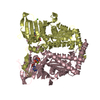
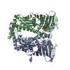
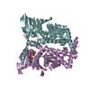
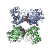
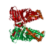
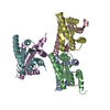
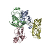
| Title | CRYSTAL STRUCTURE OF THE ATPASE REGION OF Mycobacterium tuberculosis GyrB WITH AMPPNP | ||||||
 Components Components | DNA GYRASE SUBUNIT B | ||||||
 Keywords Keywords | ISOMERASE / GHKL DOMAIN | ||||||
| Function / homology |  Function and homology information Function and homology informationDNA negative supercoiling activity / DNA topoisomerase type II (double strand cut, ATP-hydrolyzing) activity / DNA topoisomerase (ATP-hydrolysing) / DNA topological change / peptidoglycan-based cell wall / DNA-templated DNA replication / chromosome / response to antibiotic / magnesium ion binding / DNA binding ...DNA negative supercoiling activity / DNA topoisomerase type II (double strand cut, ATP-hydrolyzing) activity / DNA topoisomerase (ATP-hydrolysing) / DNA topological change / peptidoglycan-based cell wall / DNA-templated DNA replication / chromosome / response to antibiotic / magnesium ion binding / DNA binding / ATP binding / plasma membrane / cytoplasmSimilarity search - Function | ||||||
| Biological species |   MYCOBACTERIUM TUBERCULOSIS (bacteria) MYCOBACTERIUM TUBERCULOSIS (bacteria) | ||||||
| Method | X-RAY DIFFRACTION / SYNCHROTRON / MOLECULAR REPLACEMENT / Resolution: 2.95 Å | ||||||
 Authors Authors | Agrawal, A. / Roue, M. / Spitzfaden, C. / Petrella, S. / Aubry, A. / Volker, C. / Mossakowska, D. / Hann, M. / Bax, B. / Mayer, C. | ||||||
 Citation Citation | Journal: Biochem.J. / Year: 2013 Title: Mycobacterium Tuberculosis DNA Gyrase ATPase Domain Structures Suggest a Dissociative Mechanism that Explains How ATP Hydrolysis is Coupled to Domain Motion. Authors: Agrawal, A. / Roue, M. / Spitzfaden, C. / Petrella, S. / Aubry, A. / Hann, M.M. / Bax, B. / Mayer, C. | ||||||
| History |
|
- Structure visualization
| Structure viewer | Molecule: MolmilJmol/JSmol |
|---|
- Downloads & links
-Download
| PDBx/mmCIF format | 3zkd.cif.gz | 1.1 MB | Display | PDBx/mmCIF format |
|---|---|---|---|---|
| PDB format | pdb3zkd.ent.gz | 967 KB | Display | PDB format |
| PDBx/mmJSON format | 3zkd.json.gz | Tree view | PDBx/mmJSON format | |
| Others |  Other downloads Other downloads |
-Validation report
| Arichive directory | https://data.pdbj.org/pub/pdb/validation_reports/zk/3zkdftp://data.pdbj.org/pub/pdb/validation_reports/zk/3zkd | HTTPS FTP |
|---|
-Related structure data
| Related structure data |  3zkbC  3zm7C  1ei1S C: citing same article ( S: Starting model for refinement |
|---|---|
| Similar structure data |


-Links
 PDBj
PDBj


- Assembly
| Deposited unit | 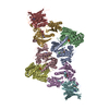
| ||||||||
|---|---|---|---|---|---|---|---|---|---|
| 1 | 
| ||||||||
| 2 | 
| ||||||||
| 3 | 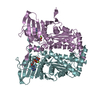
| ||||||||
| 4 | 
| ||||||||
| Unit cell |
|
-Components
| #1: Protein | Mass: 46806.246 Da / Num. of mol.: 8 / Fragment: N-TERMINAL ATPASE REGION, RESIDUES 40-466 Source method: isolated from a genetically manipulated source Details: AMPPNP IN IMINO FORM / Source: (gene. exp.) MYCOBACTERIUM TUBERCULOSIS (bacteria) / Production host: ESCHERICHIA COLI (E. coli) / Strain (production host): BL21(DE3) / Variant (production host): ROSETTAReferences: UniProt: I6WX66, UniProt: P9WG45*PLUS, EC: 5.99.1.3 #2: Chemical | ChemComp-ANP /   Mass: 506.196 Da / Num. of mol.: 8 / Source method: obtained synthetically / Formula: C10H17N6O12P3 / Comment: AMP-PNP, energy-carrying molecule analogue*YM Mass: 506.196 Da / Num. of mol.: 8 / Source method: obtained synthetically / Formula: C10H17N6O12P3 / Comment: AMP-PNP, energy-carrying molecule analogue*YM#3: Chemical | ChemComp-MG /   Mass: 24.305 Da / Num. of mol.: 8 / Source method: obtained synthetically / Formula: Mg Mass: 24.305 Da / Num. of mol.: 8 / Source method: obtained synthetically / Formula: Mg#4: Water | ChemComp-HOH / | Water Mass: 18.015 Da / Num. of mol.: 115 / Source method: isolated from a natural source / Formula: H2O Mass: 18.015 Da / Num. of mol.: 115 / Source method: isolated from a natural source / Formula: H2ONonpolymer details | AMPPNP IN IMINO FORM. THIS IS THE IMINO FORM OF AMPPNP. | Sequence details | NOTE - WE USED NEW CONSENSUS START SITE (A VALINE) WHICH IS RESIDUE 40 IN THIS ENTRY. | |
|---|
-Experimental details
-Experiment
| Experiment | Method: X-RAY DIFFRACTION / Number of used crystals: 1 |
|---|
- Sample preparation
| Crystal | Density Matthews: 2.37 Å3/Da / Density % sol: 48.01 % / Description: NONE |
|---|
-Data collection
| Diffraction | Mean temperature: 100 K |
|---|---|
| Diffraction source | Source: SYNCHROTRON / Site: ESRF  / Beamline: ID23-1 / Wavelength: 0.97955 / Beamline: ID23-1 / Wavelength: 0.97955 |
| Detector | Type: ADSC QUANTUM 315r / Detector: CCD |
| Radiation | Protocol: SINGLE WAVELENGTH / Monochromatic (M) / Laue (L): M / Scattering type: x-ray |
| Radiation wavelength | Wavelength: 0.97955 Å / Relative weight: 1 |
| Reflection | Resolution: 2.95→40 Å / Num. obs: 64548 / % possible obs: 84.5 % / Observed criterion σ(I): 0 / Redundancy: 1.68 % / Biso Wilson estimate: 66.66 Å2 / Rmerge(I) obs: 0.08 / Net I/σ(I): 10.8 |
| Reflection shell | Resolution: 2.95→3 Å / Rmerge(I) obs: 0.27 / Mean I/σ(I) obs: 1.9 / % possible all: 86.2 |
- Processing
| Software |
| |||||||||||||||||||||||||||||||||||||||||||||||||||||||||||||||||||||||||||||||||||||||||||||||||||||||||||||||||||||||||||||
|---|---|---|---|---|---|---|---|---|---|---|---|---|---|---|---|---|---|---|---|---|---|---|---|---|---|---|---|---|---|---|---|---|---|---|---|---|---|---|---|---|---|---|---|---|---|---|---|---|---|---|---|---|---|---|---|---|---|---|---|---|---|---|---|---|---|---|---|---|---|---|---|---|---|---|---|---|---|---|---|---|---|---|---|---|---|---|---|---|---|---|---|---|---|---|---|---|---|---|---|---|---|---|---|---|---|---|---|---|---|---|---|---|---|---|---|---|---|---|---|---|---|---|---|---|---|---|
| Refinement | Method to determine structure: MOLECULAR REPLACEMENT Starting model: PDB ENTRY 1EI1 Resolution: 2.95→24.81 Å / Cor.coef. Fo:Fc: 0.9079 / Cor.coef. Fo:Fc free: 0.8564 / Cross valid method: THROUGHOUT / σ(F): 0 / SU Rfree Blow DPI: 0.469
| |||||||||||||||||||||||||||||||||||||||||||||||||||||||||||||||||||||||||||||||||||||||||||||||||||||||||||||||||||||||||||||
| Displacement parameters | Biso mean: 80.27 Å2
| |||||||||||||||||||||||||||||||||||||||||||||||||||||||||||||||||||||||||||||||||||||||||||||||||||||||||||||||||||||||||||||
| Refine analyze | Luzzati coordinate error obs: 0.417 Å | |||||||||||||||||||||||||||||||||||||||||||||||||||||||||||||||||||||||||||||||||||||||||||||||||||||||||||||||||||||||||||||
| Refinement step | Cycle: LAST / Resolution: 2.95→24.81 Å
| |||||||||||||||||||||||||||||||||||||||||||||||||||||||||||||||||||||||||||||||||||||||||||||||||||||||||||||||||||||||||||||
| Refine LS restraints |
| |||||||||||||||||||||||||||||||||||||||||||||||||||||||||||||||||||||||||||||||||||||||||||||||||||||||||||||||||||||||||||||
| LS refinement shell | Resolution: 2.95→3.03 Å / Total num. of bins used: 20
| |||||||||||||||||||||||||||||||||||||||||||||||||||||||||||||||||||||||||||||||||||||||||||||||||||||||||||||||||||||||||||||
| Refinement TLS params. | Method: refined / Refine-ID: X-RAY DIFFRACTION
| |||||||||||||||||||||||||||||||||||||||||||||||||||||||||||||||||||||||||||||||||||||||||||||||||||||||||||||||||||||||||||||
| Refinement TLS group |
|
