- PDB-3zkb: CRYSTAL STRUCTURE OF THE ATPASE REGION OF Mycobacterium tuberculo... -
+
Open data
ID or keywords:
Loading...
-
Basic information
Entry
Database: PDB / ID: 3zkb
Title
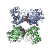
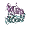
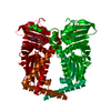
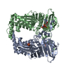
CRYSTAL STRUCTURE OF THE ATPASE REGION OF Mycobacterium tuberculosis GyrB WITH AMPPNP
Components
DNA GYRASE SUBUNIT B
Keywords
ISOMERASE / TYPE IIA TOPOISOMERASE / GHKL DOMAIN
Function / homology
Function and homology information
DNA negative supercoiling activity / DNA topoisomerase type II (double strand cut, ATP-hydrolyzing) activity / DNA topoisomerase (ATP-hydrolysing) / DNA topological change / peptidoglycan-based cell wall / DNA-templated DNA replication / chromosome / response to antibiotic / magnesium ion binding / DNA binding ...DNA negative supercoiling activity / DNA topoisomerase type II (double strand cut, ATP-hydrolyzing) activity / DNA topoisomerase (ATP-hydrolysing) / DNA topological change / peptidoglycan-based cell wall / DNA-templated DNA replication / chromosome / response to antibiotic / magnesium ion binding / DNA binding / ATP binding / plasma membrane / cytoplasm Similarity search - Function
DNA gyrase subunit B, TOPRIM domain / DNA gyrase, subunit B / DNA topoisomerase, type IIA, subunit B / DNA gyrase B subunit, C-terminal / DNA gyrase B subunit, carboxyl terminus / DNA topoisomerase, type IIA, subunit B, domain 2 / DNA gyrase B / DNA topoisomerase, type IIA / DNA topoisomerase, type IIA, conserved site / DNA topoisomerase II signature. ...DNA gyrase subunit B, TOPRIM domain / DNA gyrase, subunit B / DNA topoisomerase, type IIA, subunit B / DNA gyrase B subunit, C-terminal / DNA gyrase B subunit, carboxyl terminus / DNA topoisomerase, type IIA, subunit B, domain 2 / DNA gyrase B / DNA topoisomerase, type IIA / DNA topoisomerase, type IIA, conserved site / DNA topoisomerase II signature. / TopoisomeraseII / DNA topoisomerase, type IIA, subunit B, C-terminal / Toprim domain / DNA topoisomerase, type IIA-like domain superfamily / Ribosomal Protein S5; domain 2 - #10 / Toprim domain profile. / TOPRIM domain / Ribosomal Protein S5; domain 2 / Histidine kinase-like ATPase, C-terminal domain / Heat Shock Protein 90 / Histidine kinase-, DNA gyrase B-, and HSP90-like ATPase / Histidine kinase-like ATPases / Histidine kinase/HSP90-like ATPase / Histidine kinase/HSP90-like ATPase superfamily / Ribosomal protein S5 domain 2-type fold, subgroup / Ribosomal protein S5 domain 2-type fold / 2-Layer Sandwich / Alpha Beta Similarity search - Domain/homology
#95 - Nov 2007 Multidrug Resistance Transporters similarity (1)
-
Assembly
Deposited unit
A: DNA GYRASE SUBUNIT B B: DNA GYRASE SUBUNIT B C: DNA GYRASE SUBUNIT B D: DNA GYRASE SUBUNIT B E: DNA GYRASE SUBUNIT B F: DNA GYRASE SUBUNIT B G: DNA GYRASE SUBUNIT B H: DNA GYRASE SUBUNIT B I: DNA GYRASE SUBUNIT B J: DNA GYRASE SUBUNIT B K: DNA GYRASE SUBUNIT B L: DNA GYRASE SUBUNIT B M: DNA GYRASE SUBUNIT B N: DNA GYRASE SUBUNIT B O: DNA GYRASE SUBUNIT B P: DNA GYRASE SUBUNIT B hetero molecules
Protocol: SINGLE WAVELENGTH / Monochromatic (M) / Laue (L): M / Scattering type: x-ray
Radiation wavelength
Wavelength: 0.97855 Å / Relative weight: 1
Reflection
Resolution: 2.9→25 Å / Num. obs: 159199 / % possible obs: 98.3 % / Observed criterion σ(I): 0 / Redundancy: 1.91 % / Biso Wilson estimate: 76.57 Å2 / Rmerge(I) obs: 0.05 / Net I/σ(I): 14.1
Reflection shell
Resolution: 2.9→2.95 Å / Rmerge(I) obs: 0.42 / Mean I/σ(I) obs: 1.9 / % possible all: 97.7
-
Processing
Software
Name
Version
Classification
BUSTER
2.11.4
refinement
DENZO
datareduction
SCALEPACK
datascaling
PHASER
phasing
Refinement
Method to determine structure: MOLECULAR REPLACEMENT / Resolution: 2.9→24.94 Å / Cor.coef. Fo:Fc: 0.9171 / Cor.coef. Fo:Fc free: 0.8866 / Cross valid method: THROUGHOUT / σ(F): 0 / SU Rfree Blow DPI: 0.361 Details: IDEAL-DIST CONTACT TERM CONTACT SETUP. RESIDUE TYPES WITHOUT CCP4 ATOM TYPE IN LIBRARY=MG. NUMBER OF ATOMS WITH PROPER CCP4 ATOM TYPE=46932. NUMBER WITH APPROX DEFAULT CCP4 ATOM TYPE=0. ...Details: IDEAL-DIST CONTACT TERM CONTACT SETUP. RESIDUE TYPES WITHOUT CCP4 ATOM TYPE IN LIBRARY=MG. NUMBER OF ATOMS WITH PROPER CCP4 ATOM TYPE=46932. NUMBER WITH APPROX DEFAULT CCP4 ATOM TYPE=0. NUMBER TREATED BY BAD NON-BONDED CONTACTS=20.
Rfactor
Num. reflection
% reflection
Selection details
Rfree
0.2397
7966
5.01 %
RANDOM
Rwork
0.1816
-
-
-
obs
0.1845
159159
98.2 %
-
Displacement parameters
Biso mean: 99.07 Å2
Baniso -1
Baniso -2
Baniso -3
1-
-5.3126 Å2
-17.1504 Å2
5.0372 Å2
2-
-
25.6843 Å2
-4.7362 Å2
3-
-
-
-20.3718 Å2
Refine analyze
Luzzati coordinate error obs: 0.518 Å
Refinement step
Cycle: LAST / Resolution: 2.9→24.94 Å
Protein
Nucleic acid
Ligand
Solvent
Total
Num. atoms
45997
0
516
439
46952
Refine LS restraints
Refine-ID
Type
Dev ideal
Number
Restraint function
Weight
X-RAY DIFFRACTION
t_bond_d
0.009
47395
HARMONIC
2
X-RAY DIFFRACTION
t_angle_deg
1.1
64483
HARMONIC
2
X-RAY DIFFRACTION
t_dihedral_angle_d
16027
SINUSOIDAL
2
X-RAY DIFFRACTION
t_incorr_chiral_ct
X-RAY DIFFRACTION
t_pseud_angle
X-RAY DIFFRACTION
t_trig_c_planes
1172
HARMONIC
2
X-RAY DIFFRACTION
t_gen_planes
6985
HARMONIC
5
X-RAY DIFFRACTION
t_it
47395
HARMONIC
20
X-RAY DIFFRACTION
t_nbd
4
SEMIHARMONIC
5
X-RAY DIFFRACTION
t_omega_torsion
2.33
X-RAY DIFFRACTION
t_other_torsion
20.61
X-RAY DIFFRACTION
t_improper_torsion
X-RAY DIFFRACTION
t_chiral_improper_torsion
6344
SEMIHARMONIC
5
X-RAY DIFFRACTION
t_sum_occupancies
X-RAY DIFFRACTION
t_utility_distance
X-RAY DIFFRACTION
t_utility_angle
X-RAY DIFFRACTION
t_utility_torsion
X-RAY DIFFRACTION
t_ideal_dist_contact
54835
SEMIHARMONIC
4
LS refinement shell
Resolution: 2.9→2.98 Å / Total num. of bins used: 20
Rfactor
Num. reflection
% reflection
Rfree
0.3097
564
4.89 %
Rwork
0.2392
10968
-
all
0.2426
11532
-
obs
-
-
98.2 %
Refinement TLS params.
Method: refined / Refine-ID: X-RAY DIFFRACTION
ID
L11 (°2)
L12 (°2)
L13 (°2)
L22 (°2)
L23 (°2)
L33 (°2)
S11 (Å °)
S12 (Å °)
S13 (Å °)
S21 (Å °)
S22 (Å °)
S23 (Å °)
S31 (Å °)
S32 (Å °)
S33 (Å °)
T11 (Å2)
T12 (Å2)
T13 (Å2)
T22 (Å2)
T23 (Å2)
T33 (Å2)
Origin x (Å)
Origin y (Å)
Origin z (Å)
1
1.1828
-0.6696
0.1413
5.3968
-1.0356
1.7352
0.0301
-0.0287
0.1365
-0.3649
-0.1588
-0.3571
0.1179
0.1336
0.1287
-0.1357
0.1304
-0.0264
-0.1768
0.0146
-0.358
-19.5005
8.552
-39.3635
2
1.3997
0.2847
-0.1219
3.1182
0.0397
0.9045
-0.0845
-0.2144
-0.1051
-0.0722
-0.0308
-0.3558
0.0447
-0.0487
0.1153
-0.2386
0.1083
-0.0569
-0.1604
-0.011
-0.1171
-18.6087
-13.316
31.1052
3
1.2298
-0.6432
0.0701
2.9767
-0.7419
1.4374
0.0279
-0.1066
0.2429
0.491
0.1505
0.0023
-0.3104
0.0117
-0.1784
-0.1549
0.0425
0.0135
-0.3269
-0.0576
0.0439
-36.4011
59.6003
24.818
4
2.2034
0.1014
0.0673
2.0537
-0.294
1.8737
-0.1395
-0.0173
0.2716
-0.179
0.0152
0.0461
0.2329
0.1223
0.1243
-0.4028
0.1166
0.0394
-0.1813
-0.0998
-0.0159
-64.222
-16.2894
-6.7262
5
0.9678
-0.2749
-0.1458
3.0703
1.1271
2.7285
0.032
-0.1445
-0.1977
0.4458
0.0281
0.097
0.5145
-0.2596
-0.0602
-0.0743
-0.2093
-0.3272
-0.1101
0.168
-0.3253
-60.5893
4.5995
-77.1973
6
0.8002
-1.5889
-0.1425
4.5779
1.2329
0.9208
-0.0147
0.0059
0.1336
0.0553
-0.0593
-0.3964
-0.0653
-0.1704
0.074
-0.0978
0.1568
0.1656
-0.3231
0.0744
-0.0405
-45.7586
78.7737
-45.1336
7
0.4027
-0.2934
-0.1848
1.5964
-0.4164
2.235
0.1322
0.0137
-0.0329
-0.1469
-0.3191
-0.117
-0.2809
0.1278
0.1869
-0.3777
0.0256
0.1812
-0.1119
-0.3172
0.2078
-88.0329
55.2832
-47.9142
8
2.1225
-0.6627
-0.7875
1.5454
-0.24
0.9691
-0.0168
-0.0752
-0.9511
0.1658
-0.5083
0.2237
-0.2949
0.2129
0.5251
-0.4659
-0.1489
-0.2301
-0.454
0.0777
0.2576
-80.4933
36.1943
23.2904
Refinement TLS group
ID
Refine-ID
Refine TLS-ID
Selection details
1
X-RAY DIFFRACTION
1
CHAIN A AND RESID 12-602 AND CHAIN B AND RESID 12-602
2
X-RAY DIFFRACTION
2
CHAIN C AND RESID 12-602 AND CHAIN D AND RESID 12-602
3
X-RAY DIFFRACTION
3
CHAIN E AND RESID 12-602 AND CHAIN F AND RESID 12-602
4
X-RAY DIFFRACTION
4
CHAIN G AND RESID 12-602 AND CHAIN H AND RESID 12-602
5
X-RAY DIFFRACTION
5
CHAIN I AND RESID 12-602 AND CHAIN J AND RESID 12-602
6
X-RAY DIFFRACTION
6
CHAIN K AND RESID 12-602 AND CHAIN L AND RESID 12-602
7
X-RAY DIFFRACTION
7
CHAIN M AND RESID 12-602 AND CHAIN N AND RESID 12-602
8
X-RAY DIFFRACTION
8
CHAIN O AND RESID 12-602 AND CHAIN P AND RESID 12-602
+
About Yorodumi
-
News
-
Feb 9, 2022. New format data for meta-information of EMDB entries
New format data for meta-information of EMDB entries
Version 3 of the EMDB header file is now the official format.
The previous official version 1.9 will be removed from the archive.
In the structure databanks used in Yorodumi, some data are registered as the other names, "COVID-19 virus" and "2019-nCoV". Here are the details of the virus and the list of structure data.
Jan 31, 2019. EMDB accession codes are about to change! (news from PDBe EMDB page)
EMDB accession codes are about to change! (news from PDBe EMDB page)
The allocation of 4 digits for EMDB accession codes will soon come to an end. Whilst these codes will remain in use, new EMDB accession codes will include an additional digit and will expand incrementally as the available range of codes is exhausted. The current 4-digit format prefixed with “EMD-” (i.e. EMD-XXXX) will advance to a 5-digit format (i.e. EMD-XXXXX), and so on. It is currently estimated that the 4-digit codes will be depleted around Spring 2019, at which point the 5-digit format will come into force.
The EM Navigator/Yorodumi systems omit the EMD- prefix.
Related info.:Q: What is EMD? / ID/Accession-code notation in Yorodumi/EM Navigator
Yorodumi is a browser for structure data from EMDB, PDB, SASBDB, etc.
This page is also the successor to EM Navigator detail page, and also detail information page/front-end page for Omokage search.
The word "yorodu" (or yorozu) is an old Japanese word meaning "ten thousand". "mi" (miru) is to see.
Related info.:EMDB / PDB / SASBDB / Comparison of 3 databanks / Yorodumi Search / Aug 31, 2016. New EM Navigator & Yorodumi / Yorodumi Papers / Jmol/JSmol / Function and homology information / Changes in new EM Navigator and Yorodumi
 Movie
Movie Controller

 Yorodumi
 Open data
 Basic information
 Components
 Keywords
 Function and homology information

 Authors
 Citation
 Structure visualization
 Downloads & links
 Other downloads


 PDBj
PDBj


 Assembly


 Mass: 506.196 Da / Num. of mol.: 16 / Source method: obtained synthetically / Formula: C10H17N6O12P3 / Comment: AMP-PNP, energy-carrying molecule analogue*YM
 Mass: 24.305 Da / Num. of mol.: 20 / Source method: obtained synthetically / Formula: Mg
 Mass: 18.015 Da / Num. of mol.: 439 / Source method: isolated from a natural source / Formula: H2O
 Sample preparation
 / Beamline: ID14-4 / Wavelength: 0.97855
  Processing