 Movie
Movie Controller


+ Open data

- Basic information
| Entry | Database: PDB / ID: 3zgq | ||||||
|---|---|---|---|---|---|---|---|
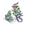
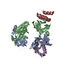
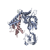
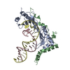
| Title | Crystal structure of human interferon-induced protein IFIT5 | ||||||
 Components Components | INTERFERON-INDUCED PROTEIN WITH TETRATRICOPEPTIDE REPEATS 5 | ||||||
 Keywords Keywords |  IMMUNE SYSTEM / RNA BINDING / INTERFERON RESPONSE IMMUNE SYSTEM / RNA BINDING / INTERFERON RESPONSE | ||||||
| Function / homology |  Function and homology informationRNA cap binding / poly(U) RNA binding / negative regulation of viral genome replication / ruffle membrane / Interferon alpha/beta signaling / actin cytoskeleton / apical part of cell / double-stranded DNA binding / positive regulation of canonical NF-kappaB signal transduction / defense response to virus ...RNA cap binding / poly(U) RNA binding / negative regulation of viral genome replication / ruffle membrane / Interferon alpha/beta signaling / actin cytoskeleton / apical part of cell / double-stranded DNA binding / positive regulation of canonical NF-kappaB signal transduction / defense response to virus / tRNA binding / single-stranded RNA binding / innate immune response / RNA binding / plasma membrane / cytosol Function and homology informationRNA cap binding / poly(U) RNA binding / negative regulation of viral genome replication / ruffle membrane / Interferon alpha/beta signaling / actin cytoskeleton / apical part of cell / double-stranded DNA binding / positive regulation of canonical NF-kappaB signal transduction / defense response to virus ...RNA cap binding / poly(U) RNA binding / negative regulation of viral genome replication / ruffle membrane / Interferon alpha/beta signaling / actin cytoskeleton / apical part of cell / double-stranded DNA binding / positive regulation of canonical NF-kappaB signal transduction / defense response to virus / tRNA binding / single-stranded RNA binding / innate immune response / RNA binding / plasma membrane / cytosolSimilarity search - Function | ||||||
| Biological species |  HOMO SAPIENS (human) HOMO SAPIENS (human) | ||||||
| Method | X-RAY DIFFRACTION / SYNCHROTRON / MAD / Resolution: 2.203 Å | ||||||
 Authors Authors | Katibah, G.E. / Lee, H.J. / Huizar, J.P. / Vogan, J.M. / Alber, T. / Collins, K. | ||||||
 Citation Citation | Journal: Mol.Cell / Year: 2013 Title: TRNA Binding, Structure, and Localization of the Human Interferon-Induced Protein Ifit5. Authors: Katibah, G.E. / Lee, H.J. / Huizar, J.P. / Vogan, J.M. / Alber, T. / Collins, K. | ||||||
| History |
|
- Structure visualization
| Structure viewer | Molecule: MolmilJmol/JSmol |
|---|
- Downloads & links
-Download
| PDBx/mmCIF format | 3zgq.cif.gz | 203.4 KB | Display | PDBx/mmCIF format |
|---|---|---|---|---|
| PDB format | pdb3zgq.ent.gz | 172 KB | Display | PDB format |
| PDBx/mmJSON format | 3zgq.json.gz | Tree view | PDBx/mmJSON format | |
| Others |  Other downloads Other downloads |
-Validation report
| Arichive directory | https://data.pdbj.org/pub/pdb/validation_reports/zg/3zgqftp://data.pdbj.org/pub/pdb/validation_reports/zg/3zgq | HTTPS FTP |
|---|
-Related structure data
| Similar structure data |
|---|


-Links
 PDBj
PDBj


- Assembly
| Deposited unit | 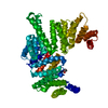
| ||||||||
|---|---|---|---|---|---|---|---|---|---|
| 1 |
| ||||||||
| Unit cell |
|
-Components
| #1: Protein | Mass: 55933.715 Da / Num. of mol.: 1 Source method: isolated from a genetically manipulated source Details: RESIDUES 190-195 ARE DISORDERED / Source: (gene. exp.) HOMO SAPIENS (human) / Production host:  ESCHERICHIA COLI (E. coli) / Strain (production host): BL21(DE3) / Variant (production host): ROSETTA / References: UniProt: Q13325 ESCHERICHIA COLI (E. coli) / Strain (production host): BL21(DE3) / Variant (production host): ROSETTA / References: UniProt: Q13325 |
|---|---|
| #2: Chemical | ChemComp-PEG / Diethylene glycol  Mass: 106.120 Da / Num. of mol.: 1 / Source method: obtained synthetically / Formula: C4H10O3 Mass: 106.120 Da / Num. of mol.: 1 / Source method: obtained synthetically / Formula: C4H10O3 |
| #3: Water | ChemComp-HOH / Water Mass: 18.015 Da / Num. of mol.: 232 / Source method: isolated from a natural source / Formula: H2O Mass: 18.015 Da / Num. of mol.: 232 / Source method: isolated from a natural source / Formula: H2O |
-Experimental details
-Experiment
| Experiment | Method: X-RAY DIFFRACTION / Number of used crystals: 1 |
|---|
- Sample preparation
| Crystal | Density Matthews: 2.39 Å3/Da / Density % sol: 48.61 % / Description: NONE |
|---|---|
| Crystal grow | pH: 6.5 / Details: pH 6.5 |
-Data collection
| Diffraction | Mean temperature: 100 K |
|---|---|
| Diffraction source | Source: SYNCHROTRON / Site: ALS  / Beamline: 8.3.1 / Wavelength: 1.1111 / Beamline: 8.3.1 / Wavelength: 1.1111 |
| Detector | Type: ADSC QUANTUM 315r / Detector: CCD / Details: DOUBLE CRYSTAL SI(111) |
| Radiation | Monochromator: DOUBLE FLAT CRYSTAL, SI(111) / Protocol: SINGLE WAVELENGTH / Monochromatic (M) / Laue (L): M / Scattering type: x-ray |
| Radiation wavelength | Wavelength: 1.1111 Å / Relative weight: 1 |
| Reflection | Resolution: 2.2→47.7 Å / Num. obs: 27780 / % possible obs: 99.9 % / Observed criterion σ(I): 2 / Redundancy: 4 % / Biso Wilson estimate: 27.28 Å2 / Rmerge(I) obs: 0.11 / Net I/σ(I): 14.6 |
| Reflection shell | Resolution: 2.2→2.24 Å / Redundancy: 4 % / Rmerge(I) obs: 0.7 / Mean I/σ(I) obs: 2 / % possible all: 100 |
- Processing
| Software |
| |||||||||||||||||||||||||||||||||||||||||||||||||||||||||||||||||||||||||||||||||||||||||||||||||||||||||||||||||||||||||||||||||||||||||||||||||||||||||||||||||||||||||||||||||||||||||||||||||||||||||||||||||||||||||||||||||
|---|---|---|---|---|---|---|---|---|---|---|---|---|---|---|---|---|---|---|---|---|---|---|---|---|---|---|---|---|---|---|---|---|---|---|---|---|---|---|---|---|---|---|---|---|---|---|---|---|---|---|---|---|---|---|---|---|---|---|---|---|---|---|---|---|---|---|---|---|---|---|---|---|---|---|---|---|---|---|---|---|---|---|---|---|---|---|---|---|---|---|---|---|---|---|---|---|---|---|---|---|---|---|---|---|---|---|---|---|---|---|---|---|---|---|---|---|---|---|---|---|---|---|---|---|---|---|---|---|---|---|---|---|---|---|---|---|---|---|---|---|---|---|---|---|---|---|---|---|---|---|---|---|---|---|---|---|---|---|---|---|---|---|---|---|---|---|---|---|---|---|---|---|---|---|---|---|---|---|---|---|---|---|---|---|---|---|---|---|---|---|---|---|---|---|---|---|---|---|---|---|---|---|---|---|---|---|---|---|---|---|---|---|---|---|---|---|---|---|---|---|---|---|---|---|---|---|
| Refinement | Method to determine structure: MAD / Resolution: 2.203→47.704 Å / SU ML: 0.26 / σ(F): 1.34 / Phase error: 23.16 / Stereochemistry target values: ML / Details: RESIDUES 190-195 ARE DISORDERED
| |||||||||||||||||||||||||||||||||||||||||||||||||||||||||||||||||||||||||||||||||||||||||||||||||||||||||||||||||||||||||||||||||||||||||||||||||||||||||||||||||||||||||||||||||||||||||||||||||||||||||||||||||||||||||||||||||
| Solvent computation | Shrinkage radii: 0.9 Å / VDW probe radii: 1.11 Å / Solvent model: FLAT BULK SOLVENT MODEL | |||||||||||||||||||||||||||||||||||||||||||||||||||||||||||||||||||||||||||||||||||||||||||||||||||||||||||||||||||||||||||||||||||||||||||||||||||||||||||||||||||||||||||||||||||||||||||||||||||||||||||||||||||||||||||||||||
| Refinement step | Cycle: LAST / Resolution: 2.203→47.704 Å
| |||||||||||||||||||||||||||||||||||||||||||||||||||||||||||||||||||||||||||||||||||||||||||||||||||||||||||||||||||||||||||||||||||||||||||||||||||||||||||||||||||||||||||||||||||||||||||||||||||||||||||||||||||||||||||||||||
| Refine LS restraints |
| |||||||||||||||||||||||||||||||||||||||||||||||||||||||||||||||||||||||||||||||||||||||||||||||||||||||||||||||||||||||||||||||||||||||||||||||||||||||||||||||||||||||||||||||||||||||||||||||||||||||||||||||||||||||||||||||||
| LS refinement shell |
| |||||||||||||||||||||||||||||||||||||||||||||||||||||||||||||||||||||||||||||||||||||||||||||||||||||||||||||||||||||||||||||||||||||||||||||||||||||||||||||||||||||||||||||||||||||||||||||||||||||||||||||||||||||||||||||||||
| Refinement TLS params. | Method: refined / Refine-ID: X-RAY DIFFRACTION
| |||||||||||||||||||||||||||||||||||||||||||||||||||||||||||||||||||||||||||||||||||||||||||||||||||||||||||||||||||||||||||||||||||||||||||||||||||||||||||||||||||||||||||||||||||||||||||||||||||||||||||||||||||||||||||||||||
| Refinement TLS group |
|
