[English] 日本語
Yorodumi
- PDB-3zew: Crystal structure of EphB4 in complex with staurosporine -

+
Open data


ID or keywords:

Loading...

-
Basic information

Entry
Database: PDB / ID: 3zew
TitleCrystal structure of EphB4 in complex with staurosporine
ComponentsEPHRIN TYPE-B RECEPTOR 4
KeywordsTRANSFERASE / INHIBITOR
Function / homology
Function and homology information


ephrin receptor activity / cell migration involved in sprouting angiogenesis / EPH-Ephrin signaling / Ephrin signaling / EPH-ephrin mediated repulsion of cells / ephrin receptor signaling pathway / heart morphogenesis / EPHB-mediated forward signaling / transmembrane receptor protein tyrosine kinase activity / receptor protein-tyrosine kinase ...ephrin receptor activity / cell migration involved in sprouting angiogenesis / EPH-Ephrin signaling / Ephrin signaling / EPH-ephrin mediated repulsion of cells / ephrin receptor signaling pathway / heart morphogenesis / EPHB-mediated forward signaling / transmembrane receptor protein tyrosine kinase activity / receptor protein-tyrosine kinase / angiogenesis / protein autophosphorylation / receptor complex / cell adhesion / extracellular exosome / extracellular region / ATP binding / plasma membrane / cytosol
Similarity search - Function
Ephrin type-B receptor 4, ligand binding domain / EPH-B4, SAM domain / Tyrosine-protein kinase ephrin type A/B receptor-like / Tyrosine-protein kinase ephrin type A/B receptor-like / Ephrin receptor type-A /type-B / Ephrin receptor ligand binding domain / Tyrosine-protein kinase, receptor class V, conserved site / Ephrin receptor, transmembrane domain / Ephrin receptor ligand binding domain / Ephrin type-A receptor 2 transmembrane domain ...Ephrin type-B receptor 4, ligand binding domain / EPH-B4, SAM domain / Tyrosine-protein kinase ephrin type A/B receptor-like / Tyrosine-protein kinase ephrin type A/B receptor-like / Ephrin receptor type-A /type-B / Ephrin receptor ligand binding domain / Tyrosine-protein kinase, receptor class V, conserved site / Ephrin receptor, transmembrane domain / Ephrin receptor ligand binding domain / Ephrin type-A receptor 2 transmembrane domain / Receptor tyrosine kinase class V signature 1. / Receptor tyrosine kinase class V signature 2. / Eph receptor ligand-binding domain profile. / Ephrin receptor ligand binding domain / Putative ephrin-receptor like / SAM domain (Sterile alpha motif) / SAM domain profile. / Sterile alpha motif. / Sterile alpha motif domain / Sterile alpha motif/pointed domain superfamily / Growth factor receptor cysteine-rich domain superfamily / EGF-like domain signature 2. / Fibronectin type III domain / Fibronectin type 3 domain / Fibronectin type-III domain profile. / Galactose-binding-like domain superfamily / Fibronectin type III / Fibronectin type III superfamily / Tyrosine-protein kinase, catalytic domain / Tyrosine kinase, catalytic domain / Tyrosine protein kinases specific active-site signature. / Tyrosine-protein kinase, active site / Protein tyrosine and serine/threonine kinase / Serine-threonine/tyrosine-protein kinase, catalytic domain / Transferase(Phosphotransferase) domain 1 / Transferase(Phosphotransferase); domain 1 / Phosphorylase Kinase; domain 1 / Phosphorylase Kinase; domain 1 / Protein kinase, ATP binding site / Protein kinases ATP-binding region signature. / Immunoglobulin-like fold / Protein kinase domain profile. / Protein kinase domain / Protein kinase-like domain superfamily / 2-Layer Sandwich / Orthogonal Bundle / Mainly Alpha / Alpha Beta
Similarity search - Domain/homology
STAUROSPORINE / Ephrin type-B receptor 4
Similarity search - Component
Biological speciesHOMO SAPIENS (human)
MethodX-RAY DIFFRACTION / MOLECULAR REPLACEMENT / Resolution: 2.5 Å
AuthorsDebreczeni, J.E. / Overman, R. / Truman, C. / McAlister, M. / Attwood, T.K.
CitationJournal: Protein Sci. / Year: 2014
Title: Completing the Structural Family Portrait of the Human Ephb Tyrosine Kinase Domains
Authors: Overman, R.C. / Debreczeni, J.E. / Truman, C.M. / Mcalister, M.S. / Attwood, T.K.
History
DepositionDec 7, 2012Deposition site: PDBE / Processing site: PDBE
Revision 1.0Dec 25, 2013Provider: repository / Type: Initial release
Revision 1.1Mar 5, 2014Group: Database references
Revision 1.2Apr 9, 2014Group: Database references
Revision 1.3May 7, 2014Group: Database references
Revision 1.4Jun 28, 2017Group: Data collection / Category: diffrn_source / Item: _diffrn_source.type

-
Structure visualization

Structure viewerMolecule:
MolmilJmol/JSmol

Downloads & links

-
Assembly

Deposited unit
A: EPHRIN TYPE-B RECEPTOR 4
B: EPHRIN TYPE-B RECEPTOR 4
hetero molecules


Theoretical massNumber of molelcules
Total (without water)68,5248
Polymers67,2072
Non-polymers1,3176
Water1,982110
1
A: EPHRIN TYPE-B RECEPTOR 4
hetero molecules


Theoretical massNumber of molelcules
Total (without water)34,2624
Polymers33,6031
Non-polymers6593
Water181
TypeNameSymmetry operationNumber
identity operation1_555x,y,z1
MethodPISA
2
B: EPHRIN TYPE-B RECEPTOR 4
hetero molecules


Theoretical massNumber of molelcules
Total (without water)34,2624
Polymers33,6031
Non-polymers6593
Water181
TypeNameSymmetry operationNumber
identity operation1_555x,y,z1
MethodPISA
Unit cell
Length a, b, c (Å)52.170, 88.710, 80.780
Angle α, β, γ (deg.)90.00, 103.05, 90.00
Int Tables number4
Space group name H-MP1211

-
Components

#1: Protein EPHRIN TYPE-B RECEPTOR 4 / EPHB4 / HEPATOMA TRANSMEMBRANE KINASE / TYROSINE-PROTEIN KINASE TYRO11


Mass: 33603.273 Da / Num. of mol.: 2 / Fragment: KINASE DOMAIN, RESIDUES 598-892
Source method: isolated from a genetically manipulated source
Source: (gene. exp.) HOMO SAPIENS (human) / Production host: ESCHERICHIA COLI (E. coli) / Strain (production host): BL21(DE3) / Variant (production host): STAR
References: UniProt: P54760, receptor protein-tyrosine kinase
#2: Chemical ChemComp-STU / STAUROSPORINE / Staurosporine


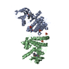
Mass: 466.531 Da / Num. of mol.: 2 / Source method: obtained synthetically / Formula: C28H26N4O3 / Comment: anticancer, antifungal, antibiotic, alkaloid*YM
#3: Chemical
ChemComp-SO4 / SULFATE ION / Sulfate


Mass: 96.063 Da / Num. of mol.: 4 / Source method: obtained synthetically / Formula: SO4
#4: Water ChemComp-HOH / water / Water


Mass: 18.015 Da / Num. of mol.: 110 / Source method: isolated from a natural source / Formula: H2O

-
Experimental details

-
Experiment

ExperimentMethod: X-RAY DIFFRACTION / Number of used crystals: 1

-
Sample preparation

CrystalDensity Matthews: 3.09 Å3/Da / Density % sol: 60.26 % / Description: NONE

-
Data collection

DiffractionMean temperature: 100 K
Diffraction sourceSource: ROTATING ANODE / Type: RIGAKU MICROMAX-007 / Wavelength: 1.5418
DetectorType: RIGAKU SATURN / Detector: CCD / Date: Jul 19, 2009 / Details: MIRRORS
RadiationMonochromator: MIRRORS / Protocol: SINGLE WAVELENGTH / Monochromatic (M) / Laue (L): M / Scattering type: x-ray
Radiation wavelengthWavelength: 1.5418 Å / Relative weight: 1
ReflectionResolution: 2.5→33.42 Å / Num. obs: 22752 / % possible obs: 91.3 % / Observed criterion σ(I): 2.4 / Redundancy: 3.22 % / Biso Wilson estimate: 65.75 Å2 / Rmerge(I) obs: 0.11 / Net I/σ(I): 6.3
Reflection shellResolution: 2.5→2.59 Å / Redundancy: 3.33 % / Rmerge(I) obs: 0.33 / Mean I/σ(I) obs: 2.4 / % possible all: 90.3

-
Processing

Software
NameVersionClassification
BUSTER2.11.5refinement
d*TREKdata reduction
DTSCALEdata scaling
PHASERphasing
RefinementMethod to determine structure: MOLECULAR REPLACEMENT / Resolution: 2.5→15 Å / Cor.coef. Fo:Fc: 0.9227 / Cor.coef. Fo:Fc free: 0.875 / SU R Cruickshank DPI: 0.532 / Cross valid method: THROUGHOUT / σ(F): 0 / SU R Blow DPI: 0.506 / SU Rfree Blow DPI: 0.306 / SU Rfree Cruickshank DPI: 0.313
Details: IDEAL-DIST CONTACT TERM CONTACT SETUP. ALL ATOMS HAVE CCP4 ATOM TYPE FROM LIBRARY
RfactorNum. reflection% reflectionSelection details
Rfree0.2816 1156 5.11 %RANDOM
Rwork0.2379 ---
obs0.2401 22618 91.21 %-
Displacement parametersBiso mean: 53.4 Å2
Baniso -1Baniso -2Baniso -3
1-0.8738 Å20 Å20.045 Å2
2---1.1673 Å20 Å2
3---0.2935 Å2
Refine analyzeLuzzati coordinate error obs: 0.438 Å
Refinement stepCycle: LAST / Resolution: 2.5→15 Å
ProteinNucleic acidLigandSolventTotal
Num. atoms4033 0 90 110 4233
Refine LS restraints
Refine-IDTypeDev idealNumberRestraint functionWeight
X-RAY DIFFRACTIONt_bond_d0.0094236HARMONIC2
X-RAY DIFFRACTIONt_angle_deg1.035774HARMONIC2
X-RAY DIFFRACTIONt_dihedral_angle_d1438SINUSOIDAL2
X-RAY DIFFRACTIONt_incorr_chiral_ct
X-RAY DIFFRACTIONt_pseud_angle
X-RAY DIFFRACTIONt_trig_c_planes99HARMONIC2
X-RAY DIFFRACTIONt_gen_planes687HARMONIC5
X-RAY DIFFRACTIONt_it4236HARMONIC20
X-RAY DIFFRACTIONt_nbd
X-RAY DIFFRACTIONt_omega_torsion2.66
X-RAY DIFFRACTIONt_other_torsion17.66
X-RAY DIFFRACTIONt_improper_torsion
X-RAY DIFFRACTIONt_chiral_improper_torsion556SEMIHARMONIC5
X-RAY DIFFRACTIONt_sum_occupancies
X-RAY DIFFRACTIONt_utility_distance
X-RAY DIFFRACTIONt_utility_angle
X-RAY DIFFRACTIONt_utility_torsion
X-RAY DIFFRACTIONt_ideal_dist_contact4961SEMIHARMONIC4
LS refinement shellResolution: 2.5→2.62 Å / Total num. of bins used: 11
RfactorNum. reflection% reflection
Rfree0.2812 150 5.07 %
Rwork0.245 2806 -
all0.2467 2956 -
obs--91.21 %

+
About Yorodumi

-
News

-
Feb 9, 2022. New format data for meta-information of EMDB entries

New format data for meta-information of EMDB entries

  • Version 3 of the EMDB header file is now the official format.
  • The previous official version 1.9 will be removed from the archive.

Related info.:EMDB header

External links:wwPDB to switch to version 3 of the EMDB data model

-
Aug 12, 2020. Covid-19 info

Covid-19 info

URL: https://pdbj.org/emnavi/covid19.php

New page: Covid-19 featured information page in EM Navigator.

Related info.:Covid-19 info / Mar 5, 2020. Novel coronavirus structure data

+
Mar 5, 2020. Novel coronavirus structure data

Novel coronavirus structure data

Related info.:Yorodumi Speices / Aug 12, 2020. Covid-19 info

External links:COVID-19 featured content - PDBj / Molecule of the Month (242):Coronavirus Proteases

+
Jan 31, 2019. EMDB accession codes are about to change! (news from PDBe EMDB page)

EMDB accession codes are about to change! (news from PDBe EMDB page)

  • The allocation of 4 digits for EMDB accession codes will soon come to an end. Whilst these codes will remain in use, new EMDB accession codes will include an additional digit and will expand incrementally as the available range of codes is exhausted. The current 4-digit format prefixed with “EMD-” (i.e. EMD-XXXX) will advance to a 5-digit format (i.e. EMD-XXXXX), and so on. It is currently estimated that the 4-digit codes will be depleted around Spring 2019, at which point the 5-digit format will come into force.
  • The EM Navigator/Yorodumi systems omit the EMD- prefix.

Related info.:Q: What is EMD? / ID/Accession-code notation in Yorodumi/EM Navigator

External links:EMDB Accession Codes are Changing Soon! / Contact to PDBj

+
Jul 12, 2017. Major update of PDB

Major update of PDB

  • wwPDB released updated PDB data conforming to the new PDBx/mmCIF dictionary.
  • This is a major update changing the version number from 4 to 5, and with Remediation, in which all the entries are updated.
  • In this update, many items about electron microscopy experimental information are reorganized (e.g. em_software).
  • Now, EM Navigator and Yorodumi are based on the updated data.

External links:wwPDB Remediation / Enriched Model Files Conforming to OneDep Data Standards Now Available in the PDB FTP Archive

-
Yorodumi

Thousand views of thousand structures

  • Yorodumi is a browser for structure data from EMDB, PDB, SASBDB, etc.
  • This page is also the successor to EM Navigator detail page, and also detail information page/front-end page for Omokage search.
  • The word "yorodu" (or yorozu) is an old Japanese word meaning "ten thousand". "mi" (miru) is to see.

Related info.:EMDB / PDB / SASBDB / Comparison of 3 databanks / Yorodumi Search / Aug 31, 2016. New EM Navigator & Yorodumi / Yorodumi Papers / Jmol/JSmol / Function and homology information / Changes in new EM Navigator and Yorodumi

Read more