 Movie
Movie Controller


+ Open data

- Basic information
| Entry | Database: PDB / ID: 3x43 | ||||||
|---|---|---|---|---|---|---|---|
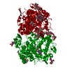
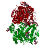
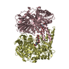
| Title | Crystal structure of O-ureido-L-serine synthase | ||||||
 Components Components | O-ureido-L-serine synthase | ||||||
 Keywords Keywords |  TRANSFERASE / D-cycloserine / type II PLP enzyme / synthase TRANSFERASE / D-cycloserine / type II PLP enzyme / synthase | ||||||
| Function / homology |  Function and homology information Function and homology informationO-ureido-L-serine synthase / cysteine synthase / cysteine synthase activity / cysteine biosynthetic process from serine / antibiotic biosynthetic processSimilarity search - Function | ||||||
| Biological species |  Streptomyces lavendulae (bacteria) Streptomyces lavendulae (bacteria) | ||||||
| Method | X-RAY DIFFRACTION / SYNCHROTRON / MOLECULAR REPLACEMENT / Resolution: 2.25 Å | ||||||
 Authors Authors | Matoba, Y. / Uda, N. / Oda, K. / Sugiyama, M. | ||||||
 Citation Citation | Journal: Febs J. / Year: 2015 Title: The structural and mutational analyses of O-ureido-L-serine synthase necessary for D-cycloserine biosynthesis. Authors: Uda, N. / Matoba, Y. / Oda, K. / Kumagai, T. / Sugiyama, M. | ||||||
| History |
|
- Structure visualization
| Structure viewer | Molecule: MolmilJmol/JSmol |
|---|
- Downloads & links
-Download
| PDBx/mmCIF format | 3x43.cif.gz | 488.4 KB | Display | PDBx/mmCIF format |
|---|---|---|---|---|
| PDB format | pdb3x43.ent.gz | 404.3 KB | Display | PDB format |
| PDBx/mmJSON format | 3x43.json.gz | Tree view | PDBx/mmJSON format | |
| Others |  Other downloads Other downloads |
-Validation report
| Arichive directory | https://data.pdbj.org/pub/pdb/validation_reports/x4/3x43ftp://data.pdbj.org/pub/pdb/validation_reports/x4/3x43 | HTTPS FTP |
|---|
-Related structure data
| Related structure data | 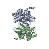 3x44C 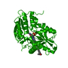 2q3dS S: Starting model for refinement C: citing same article ( |
|---|---|
| Similar structure data |


-Links
 PDBj
PDBj
- Assembly
| Deposited unit | 
| ||||||||
|---|---|---|---|---|---|---|---|---|---|
| 1 | 
| ||||||||
| 2 | 
| ||||||||
| Unit cell |
|
-Components
| #1: Protein | Mass: 35700.402 Da / Num. of mol.: 8 Source method: isolated from a genetically manipulated source Source: (gene. exp.) Streptomyces lavendulae (bacteria) / Strain: ATCC11924 / Gene: dcsD / Plasmid: pET21a(+) / Production host: Escherichia coli (E. coli) / Strain (production host): BL21(DE3)References: UniProt: D2Z027, O-ureido-L-serine synthase, cysteine synthase#2: Chemical | ChemComp-PLP / Pyridoxal phosphate  Mass: 247.142 Da / Num. of mol.: 8 / Source method: obtained synthetically / Formula: C8H10NO6P Mass: 247.142 Da / Num. of mol.: 8 / Source method: obtained synthetically / Formula: C8H10NO6P#3: Water | ChemComp-HOH / | Water Mass: 18.015 Da / Num. of mol.: 809 / Source method: isolated from a natural source / Formula: H2O Mass: 18.015 Da / Num. of mol.: 809 / Source method: isolated from a natural source / Formula: H2O |
|---|
-Experimental details
-Experiment
| Experiment | Method: X-RAY DIFFRACTION / Number of used crystals: 1 |
|---|
- Sample preparation
| Crystal | Density Matthews: 2.32 Å3/Da / Density % sol: 46.93 % |
|---|---|
| Crystal grow | Temperature: 298 K / Method: vapor diffusion, sitting drop / pH: 7.5 Details: Tris-HCl, polyethylene glycol 8,000, KH2PO4, pH 7.5, VAPOR DIFFUSION, SITTING DROP, temperature 298K |
-Data collection
| Diffraction | Mean temperature: 100 K |
|---|---|
| Diffraction source | Source: SYNCHROTRON / Site: SPring-8  / Beamline: BL41XU / Wavelength: 1 Å / Beamline: BL41XU / Wavelength: 1 Å |
| Detector | Type: RAYONIX MX-225 / Detector: CCD / Date: Dec 7, 2010 |
| Radiation | Monochromator: Rotated-inclined double-crystal / Protocol: SINGLE WAVELENGTH / Monochromatic (M) / Laue (L): M / Scattering type: x-ray |
| Radiation wavelength | Wavelength: 1 Å / Relative weight: 1 |
| Reflection | Resolution: 2.25→100 Å / Num. all: 122927 / Num. obs: 122927 / % possible obs: 99.9 % / Observed criterion σ(F): 0 / Observed criterion σ(I): 0 / Redundancy: 3.8 % / Biso Wilson estimate: 25.3 Å2 / Rmerge(I) obs: 0.09 / Net I/σ(I): 9.1 |
| Reflection shell | Resolution: 2.25→2.37 Å / Redundancy: 3.8 % / Rmerge(I) obs: 0.475 / Mean I/σ(I) obs: 2.8 / Num. unique all: 17914 / % possible all: 100 |
- Processing
| Software |
| ||||||||||||||||||||||||||||||||||||||||||||||||||||||||||||||||||||||||||||||||
|---|---|---|---|---|---|---|---|---|---|---|---|---|---|---|---|---|---|---|---|---|---|---|---|---|---|---|---|---|---|---|---|---|---|---|---|---|---|---|---|---|---|---|---|---|---|---|---|---|---|---|---|---|---|---|---|---|---|---|---|---|---|---|---|---|---|---|---|---|---|---|---|---|---|---|---|---|---|---|---|---|---|
| Refinement | Method to determine structure: MOLECULAR REPLACEMENT Starting model: 2Q3D Resolution: 2.25→29.83 Å / Rfactor Rfree error: 0.003 / Data cutoff high absF: 3018007.28 / Data cutoff low absF: 0 / Isotropic thermal model: RESTRAINED / Cross valid method: THROUGHOUT / σ(F): 2 / Stereochemistry target values: Engh & Huber / Details: BULK SOLVENT MODEL USED
| ||||||||||||||||||||||||||||||||||||||||||||||||||||||||||||||||||||||||||||||||
| Solvent computation | Solvent model: FLAT MODEL / Bsol: 46.3809 Å2 / ksol: 0.35 e/Å3 | ||||||||||||||||||||||||||||||||||||||||||||||||||||||||||||||||||||||||||||||||
| Displacement parameters | Biso mean: 51.4 Å2
| ||||||||||||||||||||||||||||||||||||||||||||||||||||||||||||||||||||||||||||||||
| Refine analyze |
| ||||||||||||||||||||||||||||||||||||||||||||||||||||||||||||||||||||||||||||||||
| Refinement step | Cycle: LAST / Resolution: 2.25→29.83 Å
| ||||||||||||||||||||||||||||||||||||||||||||||||||||||||||||||||||||||||||||||||
| Refine LS restraints |
| ||||||||||||||||||||||||||||||||||||||||||||||||||||||||||||||||||||||||||||||||
| Refine LS restraints NCS | NCS model details: NONE | ||||||||||||||||||||||||||||||||||||||||||||||||||||||||||||||||||||||||||||||||
| LS refinement shell | Resolution: 2.25→2.39 Å / Rfactor Rfree error: 0.011 / Total num. of bins used: 6
| ||||||||||||||||||||||||||||||||||||||||||||||||||||||||||||||||||||||||||||||||
| Xplor file |
|
