 Movie
Movie Controller

[English] 日本語
 Yorodumi
- PDB-3wt1: Crystal structure of the b'-a' domain of thermophilic fungal prot... -
+ Open data

- Basic information
| Entry | Database: PDB / ID: 3wt1 | ||||||
|---|---|---|---|---|---|---|---|
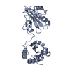
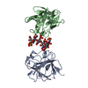
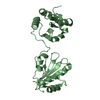
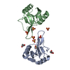
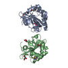
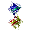
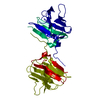
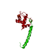
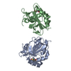
| Title | Crystal structure of the b'-a' domain of thermophilic fungal protein disulfide isomerase (reduced form) | ||||||
 Components Components | Protein disulfide-isomerase | ||||||
 Keywords Keywords | ISOMERASE / THIOREDOXIN FOLD / DISULFIDE BOND / ENDOPLASMIC RETICULUM / REDOX-ACTIVE CENTER / thioredoxin / oxidoreductase | ||||||
| Function / homology |  Function and homology informationprotein disulfide-isomerase / protein disulfide isomerase activity / response to endoplasmic reticulum stress / protein folding / endoplasmic reticulum lumen / cell surface Function and homology informationprotein disulfide-isomerase / protein disulfide isomerase activity / response to endoplasmic reticulum stress / protein folding / endoplasmic reticulum lumen / cell surfaceSimilarity search - Function | ||||||
| Biological species |  Humicola insolens (fungus) Humicola insolens (fungus) | ||||||
| Method | X-RAY DIFFRACTION / SYNCHROTRON / MOLECULAR REPLACEMENT / Resolution: 1.85 Å | ||||||
 Authors Authors | Inagaki, K. / Satoh, T. / Itoh, S.G. / Okumura, H. / Kato, K. | ||||||
 Citation Citation | Journal: Chem.Phys.Lett. / Year: 2015 Title: Redox-dependent conformational transition of catalytic domain of protein disulfide isomerase indicated by crystal structure-based molecular dynamics simulation Authors: Inagaki, K. / Satoh, T. / Itoh, S.G. / Okumura, H. / Kato, K. | ||||||
| History |
|
- Structure visualization
| Structure viewer | Molecule: MolmilJmol/JSmol |
|---|
- Downloads & links
-Download
| PDBx/mmCIF format | 3wt1.cif.gz | 198.8 KB | Display | PDBx/mmCIF format |
|---|---|---|---|---|
| PDB format | pdb3wt1.ent.gz | 159.3 KB | Display | PDB format |
| PDBx/mmJSON format | 3wt1.json.gz | Tree view | PDBx/mmJSON format | |
| Others |  Other downloads Other downloads |
-Validation report
| Arichive directory | https://data.pdbj.org/pub/pdb/validation_reports/wt/3wt1ftp://data.pdbj.org/pub/pdb/validation_reports/wt/3wt1 | HTTPS FTP |
|---|
-Related structure data
| Related structure data |  3wt2C  2kp1S S: Starting model for refinement C: citing same article ( |
|---|---|
| Similar structure data |


-Links
 PDBj
PDBj


- Assembly
| Deposited unit | 
| ||||||||||||||||||||||||||||||||||||||||||||||||||||||||||||||||||||||||||||||||||||||||||||||||||||||||||||||||||||||||||||
|---|---|---|---|---|---|---|---|---|---|---|---|---|---|---|---|---|---|---|---|---|---|---|---|---|---|---|---|---|---|---|---|---|---|---|---|---|---|---|---|---|---|---|---|---|---|---|---|---|---|---|---|---|---|---|---|---|---|---|---|---|---|---|---|---|---|---|---|---|---|---|---|---|---|---|---|---|---|---|---|---|---|---|---|---|---|---|---|---|---|---|---|---|---|---|---|---|---|---|---|---|---|---|---|---|---|---|---|---|---|---|---|---|---|---|---|---|---|---|---|---|---|---|---|---|---|
| 1 | 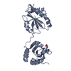
| ||||||||||||||||||||||||||||||||||||||||||||||||||||||||||||||||||||||||||||||||||||||||||||||||||||||||||||||||||||||||||||
| 2 | 
| ||||||||||||||||||||||||||||||||||||||||||||||||||||||||||||||||||||||||||||||||||||||||||||||||||||||||||||||||||||||||||||
| 3 | 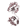
| ||||||||||||||||||||||||||||||||||||||||||||||||||||||||||||||||||||||||||||||||||||||||||||||||||||||||||||||||||||||||||||
| 4 | 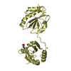
| ||||||||||||||||||||||||||||||||||||||||||||||||||||||||||||||||||||||||||||||||||||||||||||||||||||||||||||||||||||||||||||
| Unit cell |
| ||||||||||||||||||||||||||||||||||||||||||||||||||||||||||||||||||||||||||||||||||||||||||||||||||||||||||||||||||||||||||||
| Noncrystallographic symmetry (NCS) | NCS domain:
NCS domain segments: Component-ID: 0 / Beg auth comp-ID: PRO / Beg label comp-ID: PRO / End auth comp-ID: ALA / End label comp-ID: ALA / Refine code: 0
NCS ensembles :
|
-Components
| #1: Protein | / PDI Mass: 27013.658 Da / Num. of mol.: 4 / Fragment: UNP residues 278-469 Source method: isolated from a genetically manipulated source Source: (gene. exp.) Humicola insolens (fungus) / Plasmid: pGEX6P-1 / Production host:  Escherichia coli (E. coli) / Strain (production host): BL21(DE3) / References: UniProt: P55059, protein disulfide-isomerase Escherichia coli (E. coli) / Strain (production host): BL21(DE3) / References: UniProt: P55059, protein disulfide-isomerase#2: Chemical | ChemComp-GOL / Glycerol  Mass: 92.094 Da / Num. of mol.: 4 / Source method: obtained synthetically / Formula: C3H8O3 Mass: 92.094 Da / Num. of mol.: 4 / Source method: obtained synthetically / Formula: C3H8O3#3: Water | ChemComp-HOH / | Water Mass: 18.015 Da / Num. of mol.: 262 / Source method: isolated from a natural source / Formula: H2O Mass: 18.015 Da / Num. of mol.: 262 / Source method: isolated from a natural source / Formula: H2O |
|---|
-Experimental details
-Experiment
| Experiment | Method: X-RAY DIFFRACTION / Number of used crystals: 1 |
|---|
- Sample preparation
| Crystal | Density Matthews: 2.48 Å3/Da / Density % sol: 50.39 % |
|---|---|
| Crystal grow | Temperature: 293 K / Method: vapor diffusion, hanging drop / pH: 4.6 Details: 36% PEG2000MME, 100mM sodium acetate pH 4.6, 200mM ammonium sulfate, VAPOR DIFFUSION, HANGING DROP, temperature 293K |
-Data collection
| Diffraction | Mean temperature: 100 K |
|---|---|
| Diffraction source | Source: SYNCHROTRON / Site: NSRRC  / Beamline: BL13B1 / Wavelength: 1 Å / Beamline: BL13B1 / Wavelength: 1 Å |
| Detector | Type: ADSC QUANTUM 315 / Detector: CCD / Date: Aug 3, 2012 |
| Radiation | Monochromator: Si 111 / Protocol: SINGLE WAVELENGTH / Monochromatic (M) / Laue (L): M / Scattering type: x-ray |
| Radiation wavelength | Wavelength: 1 Å / Relative weight: 1 |
| Reflection | Resolution: 1.85→50 Å / Num. all: 88599 / Num. obs: 81751 / % possible obs: 92.3 % / Observed criterion σ(I): -3 / Redundancy: 2.1 % / Biso Wilson estimate: 68.4 Å2 / Rmerge(I) obs: 0.055 / Net I/σ(I): 26.2 |
| Reflection shell | Resolution: 1.85→1.88 Å / Redundancy: 2 % / Rmerge(I) obs: 0.378 / Mean I/σ(I) obs: 1.5 / Num. unique all: 3648 / % possible all: 81.7 |
- Processing
| Software |
| |||||||||||||||||||||||||||||||||||||||||||||||||||||||||||||||||
|---|---|---|---|---|---|---|---|---|---|---|---|---|---|---|---|---|---|---|---|---|---|---|---|---|---|---|---|---|---|---|---|---|---|---|---|---|---|---|---|---|---|---|---|---|---|---|---|---|---|---|---|---|---|---|---|---|---|---|---|---|---|---|---|---|---|---|
| Refinement | Method to determine structure: MOLECULAR REPLACEMENT Starting model: 2KP1 Resolution: 1.85→20 Å / Cor.coef. Fo:Fc: 0.954 / Cor.coef. Fo:Fc free: 0.937 / SU B: 6.001 / SU ML: 0.166 / Cross valid method: THROUGHOUT / ESU R: 0.189 / ESU R Free: 0.171 / Stereochemistry target values: MAXIMUM LIKELIHOOD / Details: HYDROGENS HAVE BEEN ADDED IN THE RIDING POSITIONS
| |||||||||||||||||||||||||||||||||||||||||||||||||||||||||||||||||
| Solvent computation | Ion probe radii: 0.8 Å / Shrinkage radii: 0.8 Å / VDW probe radii: 1.2 Å / Solvent model: MASK | |||||||||||||||||||||||||||||||||||||||||||||||||||||||||||||||||
| Displacement parameters | Biso mean: 41.453 Å2
| |||||||||||||||||||||||||||||||||||||||||||||||||||||||||||||||||
| Refinement step | Cycle: LAST / Resolution: 1.85→20 Å
| |||||||||||||||||||||||||||||||||||||||||||||||||||||||||||||||||
| Refine LS restraints |
| |||||||||||||||||||||||||||||||||||||||||||||||||||||||||||||||||
| Refine LS restraints NCS | Refine-ID: X-RAY DIFFRACTION / Type: interatomic distance / Weight position: 0.05
| |||||||||||||||||||||||||||||||||||||||||||||||||||||||||||||||||
| LS refinement shell | Resolution: 1.85→1.898 Å / Total num. of bins used: 20
|
