 Movie
Movie Controller

[English] 日本語
 Yorodumi
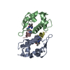
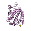
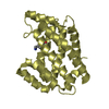
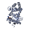
- PDB-3whp: Crystal structure of the C-terminal domain of Themus thermophilus... -
+ Open data

- Basic information
| Entry | Database: PDB / ID: 3whp | ||||||
|---|---|---|---|---|---|---|---|
| Title | Crystal structure of the C-terminal domain of Themus thermophilus LitR in complex with cobalamin | ||||||
 Components Components | Probable transcriptional regulator | ||||||
 Keywords Keywords |  GENE REGULATION / B12-binding domain / Rossmann fold / Four helix bundle / Transcriptional Regulator / Cobalamin GENE REGULATION / B12-binding domain / Rossmann fold / Four helix bundle / Transcriptional Regulator / Cobalamin | ||||||
| Function / homology |  Function and homology informationcobalamin binding / DNA-binding transcription factor activity / DNA binding / identical protein binding / metal ion binding Function and homology informationcobalamin binding / DNA-binding transcription factor activity / DNA binding / identical protein binding / metal ion bindingSimilarity search - Function | ||||||
| Biological species |   Thermus thermophilus (bacteria) Thermus thermophilus (bacteria) | ||||||
| Method | X-RAY DIFFRACTION / SYNCHROTRON / MAD / Resolution: 2.52 Å | ||||||
 Authors Authors | Agari, Y. / Takano, H. / Beppu, T. / Ueda, K. / Shinkai, A. | ||||||
 Citation Citation | Journal: To be Published Title: Crystal structure of the C-terminal domain of Themus thermophilus LitR in complex with cobalamin Authors: Agari, Y. / Takano, H. / Beppu, T. / Ueda, K. / Shinkai, A. | ||||||
| History |
|
- Structure visualization
| Structure viewer | Molecule: MolmilJmol/JSmol |
|---|
- Downloads & links
-Download
| PDBx/mmCIF format | 3whp.cif.gz | 55.5 KB | Display | PDBx/mmCIF format |
|---|---|---|---|---|
| PDB format | pdb3whp.ent.gz | 37.9 KB | Display | PDB format |
| PDBx/mmJSON format | 3whp.json.gz | Tree view | PDBx/mmJSON format | |
| Others |  Other downloads Other downloads |
-Validation report
| Arichive directory | https://data.pdbj.org/pub/pdb/validation_reports/wh/3whpftp://data.pdbj.org/pub/pdb/validation_reports/wh/3whp | HTTPS FTP |
|---|
-Related structure data
| Similar structure data |
|---|


-Links
 PDBj
PDBj

- Assembly
| Deposited unit | 
| ||||||||
|---|---|---|---|---|---|---|---|---|---|
| 1 |
| ||||||||
| Unit cell |
|
-Components
| #1: Protein | Mass: 31563.350 Da / Num. of mol.: 1 Source method: isolated from a genetically manipulated source Source: (gene. exp.) Thermus thermophilus (bacteria) / Strain: HB27 / Gene: litR, TT_P0056 / Production host: Escherichia coli (E. coli) / References: UniProt: Q746J7 |
|---|---|
| #2: Chemical | ChemComp-B12 / Vitamin B12  Mass: 1330.356 Da / Num. of mol.: 1 / Source method: obtained synthetically / Formula: C62H89CoN13O14P Mass: 1330.356 Da / Num. of mol.: 1 / Source method: obtained synthetically / Formula: C62H89CoN13O14P |
| #3: Water | ChemComp-HOH / Water Mass: 18.015 Da / Num. of mol.: 27 / Source method: isolated from a natural source / Formula: H2O Mass: 18.015 Da / Num. of mol.: 27 / Source method: isolated from a natural source / Formula: H2O |
| Nonpolymer details | THE C19 (B12 A 800) IN THIS COORDINATE |
-Experimental details
-Experiment
| Experiment | Method: X-RAY DIFFRACTION / Number of used crystals: 1 |
|---|
- Sample preparation
| Crystal | Density Matthews: 1.81 Å3/Da / Density % sol: 32.15 % |
|---|---|
| Crystal grow | Temperature: 293 K / Method: vapor diffusion, sitting drop / pH: 4.2 Details: 0.1M Phosphate-citrate, 5% PEG 1000, 35%-40% Ethanol, 0.18-0.36M Mg sulfate, 0.01-0.02M Na acetate, pH 4.2, VAPOR DIFFUSION, SITTING DROP, temperature 293K |
-Data collection
| Diffraction | Mean temperature: 100 K | |||||||||||||||
|---|---|---|---|---|---|---|---|---|---|---|---|---|---|---|---|---|
| Diffraction source | Source: SYNCHROTRON / Site: SPring-8  / Beamline: BL26B2 / Wavelength: 1.00, 1.6042, 1.6083, 1.5600 / Beamline: BL26B2 / Wavelength: 1.00, 1.6042, 1.6083, 1.5600 | |||||||||||||||
| Detector | Type: RAYONIX MX-225 / Detector: CCD / Date: Jun 27, 2012 | |||||||||||||||
| Radiation | Monochromator: A fixed exit Si double crystal monochromator followed by a two dimensional focusing mirror Protocol: MAD / Monochromatic (M) / Laue (L): M / Scattering type: x-ray | |||||||||||||||
| Radiation wavelength |
| |||||||||||||||
| Reflection | Resolution: 2.52→50 Å / Num. obs: 8257 / % possible obs: 99.8 % / Redundancy: 13 % / Biso Wilson estimate: 47.6 Å2 / Rmerge(I) obs: 0.046 / Net I/σ(I): 51.4 | |||||||||||||||
| Reflection shell | Resolution: 2.52→2.58 Å / Redundancy: 12.7 % / Rmerge(I) obs: 0.353 / Mean I/σ(I) obs: 7.8 / Num. unique all: 559 / % possible all: 100 |
- Processing
| Software |
| ||||||||||||||||||||||||||||||||||||||||||||||||||||||||||||||||||||||||||||||||
|---|---|---|---|---|---|---|---|---|---|---|---|---|---|---|---|---|---|---|---|---|---|---|---|---|---|---|---|---|---|---|---|---|---|---|---|---|---|---|---|---|---|---|---|---|---|---|---|---|---|---|---|---|---|---|---|---|---|---|---|---|---|---|---|---|---|---|---|---|---|---|---|---|---|---|---|---|---|---|---|---|---|
| Refinement | Method to determine structure: MAD / Resolution: 2.52→32.54 Å / Rfactor Rfree error: 0.008 / Data cutoff high absF: 2368756.57 / Data cutoff low absF: 0 / Isotropic thermal model: RESTRAINED / Cross valid method: THROUGHOUT / σ(F): 0 / Stereochemistry target values: Engh & Huber / Details: BULK SOLVENT MODEL USED
| ||||||||||||||||||||||||||||||||||||||||||||||||||||||||||||||||||||||||||||||||
| Solvent computation | Solvent model: FLAT MODEL / Bsol: 62.1639 Å2 / ksol: 0.35 e/Å3 | ||||||||||||||||||||||||||||||||||||||||||||||||||||||||||||||||||||||||||||||||
| Displacement parameters | Biso mean: 59.6 Å2
| ||||||||||||||||||||||||||||||||||||||||||||||||||||||||||||||||||||||||||||||||
| Refine analyze |
| ||||||||||||||||||||||||||||||||||||||||||||||||||||||||||||||||||||||||||||||||
| Refinement step | Cycle: LAST / Resolution: 2.52→32.54 Å
| ||||||||||||||||||||||||||||||||||||||||||||||||||||||||||||||||||||||||||||||||
| Refine LS restraints |
| ||||||||||||||||||||||||||||||||||||||||||||||||||||||||||||||||||||||||||||||||
| LS refinement shell | Resolution: 2.52→2.68 Å / Rfactor Rfree error: 0.028 / Total num. of bins used: 6
| ||||||||||||||||||||||||||||||||||||||||||||||||||||||||||||||||||||||||||||||||
| Xplor file |
|
