 Movie
Movie Controller

[English] 日本語
 Yorodumi
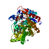
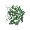
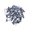
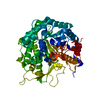
- PDB-3wbe: Rice Os3BGlu6 Beta-Glucosidase E178Q mutant in a covalent complex... -
+ Open data

- Basic information
| Entry | Database: PDB / ID: 3wbe | ||||||
|---|---|---|---|---|---|---|---|
| Title | Rice Os3BGlu6 Beta-Glucosidase E178Q mutant in a covalent complex with Glc from GA4GE. | ||||||
 Components Components | Beta-glucosidase 6 | ||||||
 Keywords Keywords | HYDROLASE / TIM BARREL / Beta-D-glucosidase / Covalently linked to alpha-D-glucoside on E394 (50% occupancy) / Secreted | ||||||
| Function / homology |  Function and homology information Function and homology informationcellobiose glucosidase activity / beta-gentiobiose beta-glucosidase activity / beta-D-fucosidase activity / glucan endo-1,3-beta-D-glucosidase activity / scopolin beta-glucosidase activity / hydrolase activity, acting on glycosyl bonds / beta-glucosidase / beta-galactosidase activity / beta-glucosidase activity / carbohydrate metabolic process / extracellular regionSimilarity search - Function | ||||||
| Biological species |  Oryza sativa Japonica Group (Japanese rice) Oryza sativa Japonica Group (Japanese rice) | ||||||
| Method | X-RAY DIFFRACTION / SYNCHROTRON / RIGID BODY REFINEMENT / Resolution: 1.97 Å | ||||||
 Authors Authors | Sansenya, S. / Hua, Y. / Cairns, J.R.K. | ||||||
 Citation Citation | Journal: Arch.Biochem.Biophys. / Year: 2013 Title: Enzymatic and structural characterization of hydrolysis of gibberellin A4 glucosyl ester by a rice beta-d-glucosidase Authors: Hua, Y. / Sansenya, S. / Saetang, C. / Wakuta, S. / Cairns, J.R.K. | ||||||
| History |
|
- Structure visualization
| Structure viewer | Molecule: MolmilJmol/JSmol |
|---|
- Downloads & links
-Download
| PDBx/mmCIF format | 3wbe.cif.gz | 124.6 KB | Display | PDBx/mmCIF format |
|---|---|---|---|---|
| PDB format | pdb3wbe.ent.gz | 94.2 KB | Display | PDB format |
| PDBx/mmJSON format | 3wbe.json.gz | Tree view | PDBx/mmJSON format | |
| Others |  Other downloads Other downloads |
-Validation report
| Arichive directory | https://data.pdbj.org/pub/pdb/validation_reports/wb/3wbeftp://data.pdbj.org/pub/pdb/validation_reports/wb/3wbe | HTTPS FTP |
|---|
-Related structure data
| Related structure data |  3wbaC  3gnoS C: citing same article ( S: Starting model for refinement |
|---|---|
| Similar structure data |


-Links
 PDBj
PDBj

- Assembly
| Deposited unit | 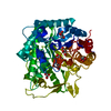
| ||||||||
|---|---|---|---|---|---|---|---|---|---|
| 1 |
| ||||||||
| Unit cell |
|
-Components
| #1: Protein | / Os3bglu6 Mass: 55373.000 Da / Num. of mol.: 1 / Fragment: Os3BGlu6 beta-glucosidase, UNP residues 38-521 / Mutation: E178Q Source method: isolated from a genetically manipulated source Details: Originally cloned into pENTR/TEV/D-TOPO and subcloned into pET32a(+)/DEST. Source: (gene. exp.) Oryza sativa Japonica Group (Japanese rice)Strain: Yukihikari / Gene: BGLU6, LOC_Os03g11420, Os03g0212800 / Plasmid: pET32a(+)/DEST / Production host:  Escherichia coli (E. coli) / Strain (production host): Origami(DE3) / References: UniProt: Q8L7J2, beta-glucosidase Escherichia coli (E. coli) / Strain (production host): Origami(DE3) / References: UniProt: Q8L7J2, beta-glucosidase | ||||
|---|---|---|---|---|---|
| #2: Sugar | ChemComp-GLC / Glucose  Type: D-saccharide, alpha linking / Mass: 180.156 Da / Num. of mol.: 1 Type: D-saccharide, alpha linking / Mass: 180.156 Da / Num. of mol.: 1Source method: isolated from a genetically manipulated source Formula: C6H12O6 | ||||
| #3: Chemical | ChemComp-GOL / Glycerol  Mass: 92.094 Da / Num. of mol.: 6 / Source method: obtained synthetically / Formula: C3H8O3 Mass: 92.094 Da / Num. of mol.: 6 / Source method: obtained synthetically / Formula: C3H8O3#4: Water | ChemComp-HOH / | Water Mass: 18.015 Da / Num. of mol.: 490 / Source method: isolated from a natural source / Formula: H2O Mass: 18.015 Da / Num. of mol.: 490 / Source method: isolated from a natural source / Formula: H2ONonpolymer details | GLC(501) AND GOL(502) ARE IN ALTERNATE CONFORMATI | |
-Experimental details
-Experiment
| Experiment | Method: X-RAY DIFFRACTION / Number of used crystals: 1 |
|---|
- Sample preparation
| Crystal | Density Matthews: 2.62 Å3/Da / Density % sol: 52.99 % |
|---|---|
| Crystal grow | Temperature: 298 K / Method: vapor diffusion, hanging drop / pH: 6.5 Details: 13% PEG5000MME, 0.1M Bis/Tris, pH 6.5, VAPOR DIFFUSION, HANGING DROP, temperature 298K |
-Data collection
| Diffraction | Mean temperature: 100 K |
|---|---|
| Diffraction source | Source: SYNCHROTRON / Site: NSRRC  / Beamline: BL13B1 / Wavelength: 1 Å / Beamline: BL13B1 / Wavelength: 1 Å |
| Detector | Type: ADSC QUANTUM 315 / Detector: CCD / Date: Sep 15, 2011 / Details: toroidal mirror |
| Radiation | Monochromator: LN2-cooled, fixed-exit double crystal monochromator, Si 111 Protocol: SINGLE WAVELENGTH / Monochromatic (M) / Laue (L): M / Scattering type: x-ray |
| Radiation wavelength | Wavelength: 1 Å / Relative weight: 1 |
| Reflection | Resolution: 1.97→30 Å / Num. all: 41777 / Num. obs: 41752 / % possible obs: 99.9 % / Redundancy: 7.1 % / Rmerge(I) obs: 0.084 / Net I/σ(I): 23.3 |
| Reflection shell | Resolution: 1.97→2.04 Å / Redundancy: 7.2 % / Rmerge(I) obs: 0.276 / Mean I/σ(I) obs: 6.6 / % possible all: 100 |
- Processing
| Software |
| ||||||||||||||||||||||||||||||||||||||||||||||||||||||||||||||||||||||||||||||||||||||||||||||||||||||||||||||||||||||||||||||||||||||||||||||||||||||||||||||||||||||||||||||||||||||
|---|---|---|---|---|---|---|---|---|---|---|---|---|---|---|---|---|---|---|---|---|---|---|---|---|---|---|---|---|---|---|---|---|---|---|---|---|---|---|---|---|---|---|---|---|---|---|---|---|---|---|---|---|---|---|---|---|---|---|---|---|---|---|---|---|---|---|---|---|---|---|---|---|---|---|---|---|---|---|---|---|---|---|---|---|---|---|---|---|---|---|---|---|---|---|---|---|---|---|---|---|---|---|---|---|---|---|---|---|---|---|---|---|---|---|---|---|---|---|---|---|---|---|---|---|---|---|---|---|---|---|---|---|---|---|---|---|---|---|---|---|---|---|---|---|---|---|---|---|---|---|---|---|---|---|---|---|---|---|---|---|---|---|---|---|---|---|---|---|---|---|---|---|---|---|---|---|---|---|---|---|---|---|---|
| Refinement | Method to determine structure: RIGID BODY REFINEMENT Starting model: PDB ENTRY 3GNO Resolution: 1.97→23.52 Å / Cor.coef. Fo:Fc: 0.968 / Cor.coef. Fo:Fc free: 0.951 / SU B: 2.392 / SU ML: 0.07 / Cross valid method: THROUGHOUT / ESU R: 0.132 / ESU R Free: 0.122 / Stereochemistry target values: MAXIMUM LIKELIHOOD / Details: HYDROGENS HAVE BEEN ADDED IN THE RIDING POSITIONS
| ||||||||||||||||||||||||||||||||||||||||||||||||||||||||||||||||||||||||||||||||||||||||||||||||||||||||||||||||||||||||||||||||||||||||||||||||||||||||||||||||||||||||||||||||||||||
| Solvent computation | Ion probe radii: 0.8 Å / Shrinkage radii: 0.8 Å / VDW probe radii: 1.4 Å / Solvent model: MASK | ||||||||||||||||||||||||||||||||||||||||||||||||||||||||||||||||||||||||||||||||||||||||||||||||||||||||||||||||||||||||||||||||||||||||||||||||||||||||||||||||||||||||||||||||||||||
| Displacement parameters | Biso mean: 22.554 Å2
| ||||||||||||||||||||||||||||||||||||||||||||||||||||||||||||||||||||||||||||||||||||||||||||||||||||||||||||||||||||||||||||||||||||||||||||||||||||||||||||||||||||||||||||||||||||||
| Refinement step | Cycle: LAST / Resolution: 1.97→23.52 Å
| ||||||||||||||||||||||||||||||||||||||||||||||||||||||||||||||||||||||||||||||||||||||||||||||||||||||||||||||||||||||||||||||||||||||||||||||||||||||||||||||||||||||||||||||||||||||
| Refine LS restraints |
|