Resolution: 1.7→45 Å / Num. obs: 62381 / % possible obs: 99.4 % / Observed criterion σ(I): 1
-
Processing
Software
Name
Version
Classification
ADSC
Quantum
datacollection
PHASER
phasing
REFMAC
5
refinement
XDSME
datareduction
XDSME
datascaling
Refinement
Method to determine structure: MOLECULAR REPLACEMENT / Resolution: 1.7→19.98 Å / Cor.coef. Fo:Fc: 0.947 / Cor.coef. Fo:Fc free: 0.924 / SU B: 2.574 / SU ML: 0.086 / Cross valid method: THROUGHOUT / ESU R: 0.133 / ESU R Free: 0.127 / Stereochemistry target values: MAXIMUM LIKELIHOOD / Details: HYDROGENS HAVE BEEN USED IF PRESENT IN THE INPUT
Rfactor
Num. reflection
% reflection
Selection details
Rfree
0.24258
3103
5.1 %
RANDOM
Rwork
0.20133
-
-
-
obs
0.20342
58159
99.91 %
-
Solvent computation
Ion probe radii: 0.8 Å / Shrinkage radii: 0.8 Å / VDW probe radii: 1.2 Å / Solvent model: MASK
 Movie
Movie Controller

 Yorodumi
 Open data
 Basic information
 Components
 Keywords
 Function and homology information

 Thermus thermophilus (bacteria)
 Authors
 Citation
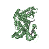
 Structure visualization
 Downloads & links
 Other downloads


 PDBj
PDBj


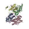
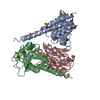
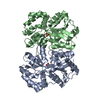
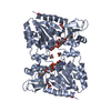
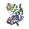
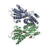
 Assembly
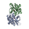

 Mass: 55.845 Da / Num. of mol.: 2 / Source method: obtained synthetically / Formula: Fe
 Mass: 61.017 Da / Num. of mol.: 4 / Source method: obtained synthetically / Formula: CHO3 / Comment: pH buffer*YM
 Mass: 18.015 Da / Num. of mol.: 513 / Source method: isolated from a natural source / Formula: H2O
 Sample preparation
 Processing