 Movie
Movie Controller


+ Open data

- Basic information
| Entry | Database: PDB / ID: 3w9t | |||||||||||||||
|---|---|---|---|---|---|---|---|---|---|---|---|---|---|---|---|---|
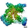
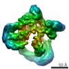
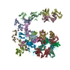
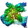
| Title | pore-forming CEL-III | |||||||||||||||
 Components Components | Hemolytic lectin CEL-III | |||||||||||||||
 Keywords Keywords |  TOXIN / Hemolytic lectin / Pore forming toxin TOXIN / Hemolytic lectin / Pore forming toxin | |||||||||||||||
| Function / homology |  Function and homology information Function and homology informationpositive regulation of erythrocyte aggregation / melibiose binding / lactose binding / cell killing / N-acetylgalactosamine binding / disruption of plasma membrane integrity in another organism / fucose binding / galactose binding / hemolysis in another organism / protein homooligomerization ...positive regulation of erythrocyte aggregation / melibiose binding / lactose binding / cell killing / N-acetylgalactosamine binding / disruption of plasma membrane integrity in another organism / fucose binding / galactose binding / hemolysis in another organism / protein homooligomerization / : / antibacterial humoral response / defense response to Gram-negative bacterium / defense response to Gram-positive bacterium / calcium ion binding / magnesium ion binding / extracellular spaceSimilarity search - Function | |||||||||||||||
| Biological species |  Cucumaria echinata (invertebrata) Cucumaria echinata (invertebrata) | |||||||||||||||
| Method | X-RAY DIFFRACTION / SYNCHROTRON / SIR / Resolution: 2.9 Å | |||||||||||||||
 Authors Authors | Unno, H. / Goda, S. / Hatakeyama, T. | |||||||||||||||
 Citation Citation | Journal: J.Biol.Chem. / Year: 2014 Title: Hemolytic lectin CEL-III heptamer reveals its transmembrane pore-formation mechanism Authors: Unno, H. / Goda, S. / Hatakeyama, T. | |||||||||||||||
| History |
|
- Structure visualization
| Structure viewer | Molecule: MolmilJmol/JSmol |
|---|
- Downloads & links
-Download
| PDBx/mmCIF format | 3w9t.cif.gz | 1.2 MB | Display | PDBx/mmCIF format |
|---|---|---|---|---|
| PDB format | pdb3w9t.ent.gz | 1019.9 KB | Display | PDB format |
| PDBx/mmJSON format | 3w9t.json.gz | Tree view | PDBx/mmJSON format | |
| Others |  Other downloads Other downloads |
-Validation report
| Arichive directory | https://data.pdbj.org/pub/pdb/validation_reports/w9/3w9tftp://data.pdbj.org/pub/pdb/validation_reports/w9/3w9t | HTTPS FTP |
|---|
-Related structure data
| Similar structure data |
|---|


-Links
 PDBj
PDBj

- Assembly
| Deposited unit | 
| ||||||||||||||||||||||||||||||||
|---|---|---|---|---|---|---|---|---|---|---|---|---|---|---|---|---|---|---|---|---|---|---|---|---|---|---|---|---|---|---|---|---|---|
| 1 |
| ||||||||||||||||||||||||||||||||
| Unit cell |
| ||||||||||||||||||||||||||||||||
| Noncrystallographic symmetry (NCS) | NCS oper:
|
-Components
| #1: Protein | Mass: 47452.164 Da / Num. of mol.: 7 / Source method: isolated from a natural source / Source: (natural) Cucumaria echinata (invertebrata) / References: UniProt: Q868M7*PLUS#2: Polysaccharide | beta-D-galactopyranose-(1-4)-beta-D-fructofuranose / lactulose /   , Oligosaccharide / Class: Water retention / Mass: 342.297 Da / Num. of mol.: 37 , Oligosaccharide / Class: Water retention / Mass: 342.297 Da / Num. of mol.: 37Source method: isolated from a genetically manipulated source Details: oligosaccharide / References: lactulose#3: Chemical | ChemComp-CA /   Mass: 40.078 Da / Num. of mol.: 44 / Source method: obtained synthetically / Formula: Ca Mass: 40.078 Da / Num. of mol.: 44 / Source method: obtained synthetically / Formula: Ca#4: Chemical | ChemComp-MG /   Mass: 24.305 Da / Num. of mol.: 14 / Source method: obtained synthetically / Formula: Mg Mass: 24.305 Da / Num. of mol.: 14 / Source method: obtained synthetically / Formula: Mg#5: Water | ChemComp-HOH / | Water Mass: 18.015 Da / Num. of mol.: 75 / Source method: isolated from a natural source / Formula: H2O Mass: 18.015 Da / Num. of mol.: 75 / Source method: isolated from a natural source / Formula: H2O |
|---|
-Experimental details
-Experiment
| Experiment | Method: X-RAY DIFFRACTION / Number of used crystals: 1 |
|---|
- Sample preparation
| Crystal | Density Matthews: 4.01 Å3/Da / Density % sol: 69.34 % |
|---|---|
| Crystal grow | Temperature: 293 K / Method: vapor diffusion / pH: 4.2 Details: 30% PEG400, 0.1M CdCl2, 0.1M sodium acetate, 10mM CaCl2, 0.1M lactulose, pH 4.2, VAPOR DIFFUSION, temperature 293K |
-Data collection
| Diffraction | Mean temperature: 95 K |
|---|---|
| Diffraction source | Source: SYNCHROTRON / Site: Diamond  / Beamline: I03 / Wavelength: 0.9763 Å / Beamline: I03 / Wavelength: 0.9763 Å |
| Detector | Type: ADSC QUANTUM 315r / Detector: CCD / Date: Oct 12, 2010 |
| Radiation | Monochromator: Si (111) double crystal monochromator / Protocol: SINGLE WAVELENGTH / Monochromatic (M) / Laue (L): M / Scattering type: x-ray |
| Radiation wavelength | Wavelength: 0.9763 Å / Relative weight: 1 |
| Reflection | Resolution: 2.9→53.5 Å / Num. obs: 113863 / % possible obs: 98.6 % / Redundancy: 3.9 % / Rmerge(I) obs: 0.1 / Net I/σ(I): 7.1 |
| Reflection shell | Resolution: 2.9→3.06 Å / Redundancy: 3.9 % / Rmerge(I) obs: 0.411 / Mean I/σ(I) obs: 2 / % possible all: 99.3 |
- Processing
| Software |
| ||||||||||||||||||||||||||||||||||||||||||||||||||||||||||||||||||||||||||||||||||||||||||||||||||||||||||||||||||||||||||||||||||||||||||||||||||||||||||||||||||||||||||||||||||||||||||||||||||||||||
|---|---|---|---|---|---|---|---|---|---|---|---|---|---|---|---|---|---|---|---|---|---|---|---|---|---|---|---|---|---|---|---|---|---|---|---|---|---|---|---|---|---|---|---|---|---|---|---|---|---|---|---|---|---|---|---|---|---|---|---|---|---|---|---|---|---|---|---|---|---|---|---|---|---|---|---|---|---|---|---|---|---|---|---|---|---|---|---|---|---|---|---|---|---|---|---|---|---|---|---|---|---|---|---|---|---|---|---|---|---|---|---|---|---|---|---|---|---|---|---|---|---|---|---|---|---|---|---|---|---|---|---|---|---|---|---|---|---|---|---|---|---|---|---|---|---|---|---|---|---|---|---|---|---|---|---|---|---|---|---|---|---|---|---|---|---|---|---|---|---|---|---|---|---|---|---|---|---|---|---|---|---|---|---|---|---|---|---|---|---|---|---|---|---|---|---|---|---|---|---|---|---|
| Refinement | Method to determine structure: SIR / Resolution: 2.9→48.2 Å / Cor.coef. Fo:Fc: 0.918 / Cor.coef. Fo:Fc free: 0.895 / SU B: 37.412 / SU ML: 0.311 / Cross valid method: THROUGHOUT / ESU R: 1.039 / ESU R Free: 0.383 / Stereochemistry target values: MAXIMUM LIKELIHOOD / Details: HYDROGENS HAVE BEEN USED IF PRESENT IN THE INPUT
| ||||||||||||||||||||||||||||||||||||||||||||||||||||||||||||||||||||||||||||||||||||||||||||||||||||||||||||||||||||||||||||||||||||||||||||||||||||||||||||||||||||||||||||||||||||||||||||||||||||||||
| Solvent computation | Ion probe radii: 0.8 Å / Shrinkage radii: 0.8 Å / VDW probe radii: 1.2 Å / Solvent model: MASK | ||||||||||||||||||||||||||||||||||||||||||||||||||||||||||||||||||||||||||||||||||||||||||||||||||||||||||||||||||||||||||||||||||||||||||||||||||||||||||||||||||||||||||||||||||||||||||||||||||||||||
| Displacement parameters | Biso mean: 80.149 Å2
| ||||||||||||||||||||||||||||||||||||||||||||||||||||||||||||||||||||||||||||||||||||||||||||||||||||||||||||||||||||||||||||||||||||||||||||||||||||||||||||||||||||||||||||||||||||||||||||||||||||||||
| Refinement step | Cycle: LAST / Resolution: 2.9→48.2 Å
| ||||||||||||||||||||||||||||||||||||||||||||||||||||||||||||||||||||||||||||||||||||||||||||||||||||||||||||||||||||||||||||||||||||||||||||||||||||||||||||||||||||||||||||||||||||||||||||||||||||||||
| Refine LS restraints |
| ||||||||||||||||||||||||||||||||||||||||||||||||||||||||||||||||||||||||||||||||||||||||||||||||||||||||||||||||||||||||||||||||||||||||||||||||||||||||||||||||||||||||||||||||||||||||||||||||||||||||
| LS refinement shell | Resolution: 2.9→2.975 Å / Total num. of bins used: 20
| ||||||||||||||||||||||||||||||||||||||||||||||||||||||||||||||||||||||||||||||||||||||||||||||||||||||||||||||||||||||||||||||||||||||||||||||||||||||||||||||||||||||||||||||||||||||||||||||||||||||||
| Refinement TLS params. | Method: refined / Refine-ID: X-RAY DIFFRACTION
| ||||||||||||||||||||||||||||||||||||||||||||||||||||||||||||||||||||||||||||||||||||||||||||||||||||||||||||||||||||||||||||||||||||||||||||||||||||||||||||||||||||||||||||||||||||||||||||||||||||||||
| Refinement TLS group |
|
