 Movie
Movie Controller

[English] 日本語
 Yorodumi
- PDB-3w8i: Crystal structure of CCM3 in complex with the C-terminal regulato... -
+ Open data

- Basic information
| Entry | Database: PDB / ID: 3w8i | ||||||
|---|---|---|---|---|---|---|---|
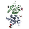
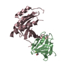
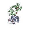
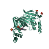
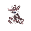
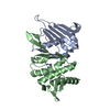
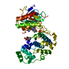
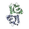
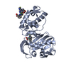
| Title | Crystal structure of CCM3 in complex with the C-terminal regulatory domain of MST4 | ||||||
 Components Components |
| ||||||
 Keywords Keywords | PROTEIN BINDING/TRANSFERASE / PROTEIN BINDING-TRANSFERASE complex | ||||||
| Function / homology |  Function and homology information Function and homology informationFAR/SIN/STRIPAK complex / intrinsic apoptotic signaling pathway in response to hydrogen peroxide / regulation of Golgi organization /  microvillus assembly / negative regulation of blood vessel endothelial cell proliferation involved in sprouting angiogenesis / Golgi reassembly / positive regulation of stress-activated MAPK cascade / endothelium development / establishment of Golgi localization / positive regulation of intracellular protein transport ...FAR/SIN/STRIPAK complex / intrinsic apoptotic signaling pathway in response to hydrogen peroxide / regulation of Golgi organization / microvillus assembly / negative regulation of blood vessel endothelial cell proliferation involved in sprouting angiogenesis / Golgi reassembly / positive regulation of stress-activated MAPK cascade / endothelium development / establishment of Golgi localization / positive regulation of intracellular protein transport / vesicle membrane / negative regulation of cell migration involved in sprouting angiogenesis / wound healing, spreading of cells / Golgi-associated vesicle / positive regulation of Notch signaling pathway / Apoptotic cleavage of cellular proteins / regulation of angiogenesis / cellular response to starvation / negative regulation of cell migration / cellular response to leukemia inhibitory factor / cell periphery / positive regulation of MAP kinase activity / positive regulation of protein serine/threonine kinase activity / positive regulation of peptidyl-serine phosphorylation / cellular response to oxidative stress / regulation of apoptotic process / angiogenesis / protein autophosphorylation / protein stabilization / non-specific serine/threonine protein kinase / protein kinase activity / intracellular signal transduction / positive regulation of cell migration / apical plasma membrane / Golgi membrane / protein phosphorylation / negative regulation of gene expression / protein serine kinase activity / protein serine/threonine kinase activity / apoptotic process / positive regulation of cell population proliferation / positive regulation of gene expression / negative regulation of apoptotic process / protein kinase binding / perinuclear region of cytoplasm / Golgi apparatus / magnesium ion binding / protein homodimerization activity / extracellular exosome / ATP binding / membrane / identical protein binding / plasma membrane / cytosol / cytoplasm microvillus assembly / negative regulation of blood vessel endothelial cell proliferation involved in sprouting angiogenesis / Golgi reassembly / positive regulation of stress-activated MAPK cascade / endothelium development / establishment of Golgi localization / positive regulation of intracellular protein transport ...FAR/SIN/STRIPAK complex / intrinsic apoptotic signaling pathway in response to hydrogen peroxide / regulation of Golgi organization / microvillus assembly / negative regulation of blood vessel endothelial cell proliferation involved in sprouting angiogenesis / Golgi reassembly / positive regulation of stress-activated MAPK cascade / endothelium development / establishment of Golgi localization / positive regulation of intracellular protein transport / vesicle membrane / negative regulation of cell migration involved in sprouting angiogenesis / wound healing, spreading of cells / Golgi-associated vesicle / positive regulation of Notch signaling pathway / Apoptotic cleavage of cellular proteins / regulation of angiogenesis / cellular response to starvation / negative regulation of cell migration / cellular response to leukemia inhibitory factor / cell periphery / positive regulation of MAP kinase activity / positive regulation of protein serine/threonine kinase activity / positive regulation of peptidyl-serine phosphorylation / cellular response to oxidative stress / regulation of apoptotic process / angiogenesis / protein autophosphorylation / protein stabilization / non-specific serine/threonine protein kinase / protein kinase activity / intracellular signal transduction / positive regulation of cell migration / apical plasma membrane / Golgi membrane / protein phosphorylation / negative regulation of gene expression / protein serine kinase activity / protein serine/threonine kinase activity / apoptotic process / positive regulation of cell population proliferation / positive regulation of gene expression / negative regulation of apoptotic process / protein kinase binding / perinuclear region of cytoplasm / Golgi apparatus / magnesium ion binding / protein homodimerization activity / extracellular exosome / ATP binding / membrane / identical protein binding / plasma membrane / cytosol / cytoplasmSimilarity search - Function | ||||||
| Biological species |  Homo sapiens (human) Homo sapiens (human) | ||||||
| Method | X-RAY DIFFRACTION / MOLECULAR REPLACEMENT / Resolution: 2.4 Å | ||||||
 Authors Authors | Xu, X. / Wang, D.C. / Ding, J. | ||||||
 Citation Citation | Journal: Structure / Year: 2013 Title: Structural Basis for the Unique Heterodimeric Assembly between Cerebral Cavernous Malformation 3 and Germinal Center Kinase III. Authors: Xu, X. / Wang, X. / Zhang, Y. / Wang, D.C. / Ding, J. | ||||||
| History |
|
- Structure visualization
| Structure viewer | Molecule: MolmilJmol/JSmol |
|---|
- Downloads & links
-Download
| PDBx/mmCIF format | 3w8i.cif.gz | 65.2 KB | Display | PDBx/mmCIF format |
|---|---|---|---|---|
| PDB format | pdb3w8i.ent.gz | 47.5 KB | Display | PDB format |
| PDBx/mmJSON format | 3w8i.json.gz | Tree view | PDBx/mmJSON format | |
| Others |  Other downloads Other downloads |
-Validation report
| Arichive directory | https://data.pdbj.org/pub/pdb/validation_reports/w8/3w8iftp://data.pdbj.org/pub/pdb/validation_reports/w8/3w8i | HTTPS FTP |
|---|
-Related structure data
| Related structure data | 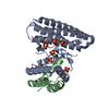 3w8hC 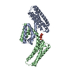 3ajmS C: citing same article ( S: Starting model for refinement |
|---|---|
| Similar structure data |

-Links
 PDBj
PDBj
- Assembly
| Deposited unit | 
| ||||||||
|---|---|---|---|---|---|---|---|---|---|
| 1 |
| ||||||||
| Unit cell |
|
-Components
| #1: Protein | / Cerebral cavernous malformations 3 protein / TF-1 cell apoptosis-related protein 15 Mass: 24896.619 Da / Num. of mol.: 1 / Fragment: UNP RESIDUES 8-212 Source method: isolated from a genetically manipulated source Source: (gene. exp.) Homo sapiens (human) / Gene: CCM3, PDCD10, TFAR15 / Plasmid: pET22b / Production host:  Escherichia coli (E. coli) / Strain (production host): BL21(DE3) / References: UniProt: Q9BUL8 Escherichia coli (E. coli) / Strain (production host): BL21(DE3) / References: UniProt: Q9BUL8 |
|---|---|
| #2: Protein | Mass: 8205.410 Da / Num. of mol.: 1 / Fragment: UNP RESIDUES 346-416 Source method: isolated from a genetically manipulated source Source: (gene. exp.) Homo sapiens (human) / Gene: MASK, MST4, SOK1, STK25, YSK1 / Plasmid: pET32a / Production host: Escherichia coli (E. coli) / Strain (production host): BL21(DE3)References: UniProt: Q9P289, non-specific serine/threonine protein kinase |
| #3: Water | ChemComp-HOH / Water Mass: 18.015 Da / Num. of mol.: 112 / Source method: isolated from a natural source / Formula: H2O Mass: 18.015 Da / Num. of mol.: 112 / Source method: isolated from a natural source / Formula: H2O |
-Experimental details
-Experiment
| Experiment | Method: X-RAY DIFFRACTION / Number of used crystals: 1 |
|---|
- Sample preparation
| Crystal | Density Matthews: 2.12 Å3/Da / Density % sol: 41.98 % |
|---|---|
| Crystal grow | Temperature: 293 K / Method: vapor diffusion, sitting drop / pH: 6 Details: 0.1M Bis-Tris, 25% PEG3350, 0.3M ammonium acetate, pH 6.0, VAPOR DIFFUSION, SITTING DROP, temperature 293K |
-Data collection
| Diffraction | Mean temperature: 95 K |
|---|---|
| Diffraction source | Source: ROTATING ANODE / Type: RIGAKU FR-E+ DW / Wavelength: 1.5418 Å |
| Detector | Type: RIGAKU RAXIS IV++ / Detector: IMAGE PLATE / Date: Nov 3, 2011 |
| Radiation | Protocol: SINGLE WAVELENGTH / Monochromatic (M) / Laue (L): M / Scattering type: x-ray |
| Radiation wavelength | Wavelength: 1.5418 Å / Relative weight: 1 |
| Reflection | Resolution: 2.4→48.85 Å / Num. all: 11764 / Num. obs: 11764 / % possible obs: 100 % / Observed criterion σ(F): 0 / Observed criterion σ(I): 0 / Redundancy: 12.8 % / Biso Wilson estimate: 37.57 Å2 / Rmerge(I) obs: 0.076 / Rsym value: 0.076 / Net I/σ(I): 27.3 |
| Reflection shell | Resolution: 2.4→2.53 Å / Redundancy: 12.7 % / Rmerge(I) obs: 0.353 / Mean I/σ(I) obs: 7.4 / Num. unique all: 1666 / Rsym value: 0.353 / % possible all: 100 |
- Processing
| Software |
| |||||||||||||||||||||||||
|---|---|---|---|---|---|---|---|---|---|---|---|---|---|---|---|---|---|---|---|---|---|---|---|---|---|---|
| Refinement | Method to determine structure: MOLECULAR REPLACEMENT Starting model: PDB ENTRY 3AJM Resolution: 2.4→45.12 Å / SU ML: 0.27 / σ(F): 1.35 / Phase error: 26.3 / Stereochemistry target values: ML
| |||||||||||||||||||||||||
| Solvent computation | Shrinkage radii: 0.6 Å / VDW probe radii: 0.9 Å / Solvent model: FLAT BULK SOLVENT MODEL / Bsol: 30.618 Å2 / ksol: 0.328 e/Å3 | |||||||||||||||||||||||||
| Displacement parameters |
| |||||||||||||||||||||||||
| Refine analyze | Luzzati coordinate error obs: 0.27 Å | |||||||||||||||||||||||||
| Refinement step | Cycle: LAST / Resolution: 2.4→45.12 Å
| |||||||||||||||||||||||||
| Refine LS restraints |
| |||||||||||||||||||||||||
| LS refinement shell | Refine-ID: X-RAY DIFFRACTION / Total num. of bins used: 4 / % reflection obs: 100 %
|
