 Movie
Movie Controller

[English] 日本語
 Yorodumi
- PDB-3w3u: Crystal structure of Kap121p mutant R349A/Q350A/D353A/E396A/N430K... -
+ Open data

- Basic information
| Entry | Database: PDB / ID: 3w3u | ||||||
|---|---|---|---|---|---|---|---|
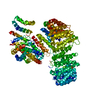
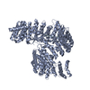
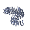
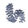
| Title | Crystal structure of Kap121p mutant R349A/Q350A/D353A/E396A/N430K/D438A/N477A | ||||||
 Components Components | Importin subunit beta-3 | ||||||
 Keywords Keywords | PROTEIN TRANSPORT / HEAT repeat / nuclear import | ||||||
| Function / homology |  Function and homology information Function and homology informationregulation of protein desumoylation / nuclear import signal receptor activity / nuclear localization sequence binding / regulation of mitotic nuclear division / mRNA export from nucleus / protein import into nucleus / nucleus / cytoplasmSimilarity search - Function | ||||||
| Biological species |  Saccharomyces cerevisiae (brewer's yeast) Saccharomyces cerevisiae (brewer's yeast) | ||||||
| Method | X-RAY DIFFRACTION / SYNCHROTRON / MOLECULAR REPLACEMENT / Resolution: 2.6 Å | ||||||
 Authors Authors | Kobayashi, J. / Matsuura, Y. | ||||||
 Citation Citation | Journal: J.Mol.Biol. / Year: 2013 Title: Structural basis for cell-cycle-dependent nuclear import mediated by the karyopherin Kap121p. Authors: Kobayashi, J. / Matsuura, Y. | ||||||
| History |
|
- Structure visualization
| Structure viewer | Molecule: MolmilJmol/JSmol |
|---|
- Downloads & links
-Download
| PDBx/mmCIF format | 3w3u.cif.gz | 209 KB | Display | PDBx/mmCIF format |
|---|---|---|---|---|
| PDB format | pdb3w3u.ent.gz | 162.4 KB | Display | PDB format |
| PDBx/mmJSON format | 3w3u.json.gz | Tree view | PDBx/mmJSON format | |
| Others |  Other downloads Other downloads |
-Validation report
| Arichive directory | https://data.pdbj.org/pub/pdb/validation_reports/w3/3w3uftp://data.pdbj.org/pub/pdb/validation_reports/w3/3w3u | HTTPS FTP |
|---|
-Related structure data
| Related structure data | 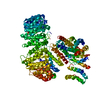 3w3tSC  3w3vC 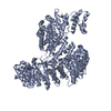 3w3wC  3w3xC  3w3yC 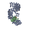 3w3zC S: Starting model for refinement C: citing same article ( |
|---|---|
| Similar structure data |


-Links
 PDBj
PDBj
- Assembly
| Deposited unit | 
| ||||||||
|---|---|---|---|---|---|---|---|---|---|
| 1 |
| ||||||||
| Unit cell |
|
-Components
| #1: Protein | / Karyopherin subunit beta-3 / Karyopherin-121 / Protein secretion enhancer 1 Mass: 119595.180 Da / Num. of mol.: 1 / Mutation: R349A, Q350A, D353A, E396A, N430K, D438A, N477A Source method: isolated from a genetically manipulated source Source: (gene. exp.) Saccharomyces cerevisiae (brewer's yeast)Strain: ATCC 204508 / S288c / Gene: PSE1, KAP121, YMR308C, YM9952.10C / Production host:  Escherichia coli (E. coli) / References: UniProt: P32337 Escherichia coli (E. coli) / References: UniProt: P32337 |
|---|
-Experimental details
-Experiment
| Experiment | Method: X-RAY DIFFRACTION / Number of used crystals: 1 |
|---|
- Sample preparation
| Crystal | Density Matthews: 3.05 Å3/Da / Density % sol: 59.69 % |
|---|---|
| Crystal grow | Temperature: 293 K / Method: vapor diffusion, hanging drop / pH: 7 Details: 0.1M HEPES, 10% 2-propanol, 24% PEG 20000, pH 7.0, VAPOR DIFFUSION, HANGING DROP, temperature 293K |
-Data collection
| Diffraction | Mean temperature: 100 K |
|---|---|
| Diffraction source | Source: SYNCHROTRON / Site: SPring-8  / Beamline: BL41XU / Wavelength: 1 Å / Beamline: BL41XU / Wavelength: 1 Å |
| Detector | Type: RAYONIX MX225HE / Detector: CCD / Date: Dec 14, 2011 |
| Radiation | Monochromator: rotated-inclined double-crystal monochromator Protocol: SINGLE WAVELENGTH / Monochromatic (M) / Laue (L): M / Scattering type: x-ray |
| Radiation wavelength | Wavelength: 1 Å / Relative weight: 1 |
| Reflection | Resolution: 2.6→26.55 Å / Num. all: 44098 / Num. obs: 43084 / % possible obs: 97.7 % / Observed criterion σ(F): 0 / Observed criterion σ(I): 0 |
| Reflection shell | Resolution: 2.6→2.7 Å / % possible all: 84.3 |
- Processing
| Software |
| ||||||||||||||||||||||||||||||||||||||||||||||||||||||||||||||||||||||||||||||||||||||||||||||||||||||||||||||||||||||||||||||||||||||||||||||||||||||||||||||||||||||||||
|---|---|---|---|---|---|---|---|---|---|---|---|---|---|---|---|---|---|---|---|---|---|---|---|---|---|---|---|---|---|---|---|---|---|---|---|---|---|---|---|---|---|---|---|---|---|---|---|---|---|---|---|---|---|---|---|---|---|---|---|---|---|---|---|---|---|---|---|---|---|---|---|---|---|---|---|---|---|---|---|---|---|---|---|---|---|---|---|---|---|---|---|---|---|---|---|---|---|---|---|---|---|---|---|---|---|---|---|---|---|---|---|---|---|---|---|---|---|---|---|---|---|---|---|---|---|---|---|---|---|---|---|---|---|---|---|---|---|---|---|---|---|---|---|---|---|---|---|---|---|---|---|---|---|---|---|---|---|---|---|---|---|---|---|---|---|---|---|---|---|---|---|
| Refinement | Method to determine structure: MOLECULAR REPLACEMENT Starting model: PDB entry 3W3T Resolution: 2.6→26.54 Å / Cor.coef. Fo:Fc: 0.946 / Cor.coef. Fo:Fc free: 0.921 / SU B: 12.213 / SU ML: 0.257 / Cross valid method: THROUGHOUT / ESU R: 0.537 / ESU R Free: 0.312 / Stereochemistry target values: MAXIMUM LIKELIHOOD / Details: HYDROGENS HAVE BEEN USED IF PRESENT IN THE INPUT
| ||||||||||||||||||||||||||||||||||||||||||||||||||||||||||||||||||||||||||||||||||||||||||||||||||||||||||||||||||||||||||||||||||||||||||||||||||||||||||||||||||||||||||
| Solvent computation | Ion probe radii: 0.8 Å / Shrinkage radii: 0.8 Å / VDW probe radii: 1.2 Å / Solvent model: MASK | ||||||||||||||||||||||||||||||||||||||||||||||||||||||||||||||||||||||||||||||||||||||||||||||||||||||||||||||||||||||||||||||||||||||||||||||||||||||||||||||||||||||||||
| Displacement parameters | Biso mean: 74.719 Å2
| ||||||||||||||||||||||||||||||||||||||||||||||||||||||||||||||||||||||||||||||||||||||||||||||||||||||||||||||||||||||||||||||||||||||||||||||||||||||||||||||||||||||||||
| Refinement step | Cycle: LAST / Resolution: 2.6→26.54 Å
| ||||||||||||||||||||||||||||||||||||||||||||||||||||||||||||||||||||||||||||||||||||||||||||||||||||||||||||||||||||||||||||||||||||||||||||||||||||||||||||||||||||||||||
| Refine LS restraints |
| ||||||||||||||||||||||||||||||||||||||||||||||||||||||||||||||||||||||||||||||||||||||||||||||||||||||||||||||||||||||||||||||||||||||||||||||||||||||||||||||||||||||||||
| LS refinement shell | Resolution: 2.6→2.668 Å / Total num. of bins used: 20
|
