 Movie
Movie Controller

[English] 日本語
 Yorodumi
- PDB-3vuu: Crystal structure of the merozoite surface protein MSPDBL2 from P... -
+ Open data

- Basic information
| Entry | Database: PDB / ID: 3vuu | ||||||
|---|---|---|---|---|---|---|---|
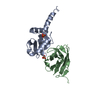
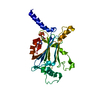
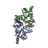
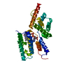
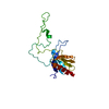
| Title | Crystal structure of the merozoite surface protein MSPDBL2 from P. falciparum | ||||||
 Components Components | Erythrocyte membrane protein, putative Red blood cell Red blood cell | ||||||
 Keywords Keywords | CELL ADHESION / Duffy Binding-Like domain / Erythrocyte Binding / Merozoite Surface / Malaria | ||||||
| Function / homology |  Function and homology information Function and homology informationhost cell surface binding / host cell surface receptor binding / cell surface / membrane / metal ion bindingSimilarity search - Function | ||||||
| Biological species |  Plasmodium falciparum (malaria parasite P. falciparum) Plasmodium falciparum (malaria parasite P. falciparum) | ||||||
| Method | X-RAY DIFFRACTION / SYNCHROTRON / MOLECULAR REPLACEMENT / Resolution: 2.093 Å | ||||||
 Authors Authors | Czabotar, P.E. / Hodder, A.N. / Clarke, O.B. / Lin, C.S. / Smith, B.J. / Cowman, A.F. | ||||||
 Citation Citation | Journal: J.Biol.Chem. / Year: 2012 Title: Insights into Duffy binding-like domains through the crystal structure and function of the merozoite surface protein MSPDBL2 from Plasmodium falciparum Authors: Hodder, A.N. / Czabotar, P.E. / Uboldi, A.D. / Clarke, O.B. / Lin, C.S. / Healer, J. / Smith, B.J. / Cowman, A.F. | ||||||
| History |
|
- Structure visualization
| Structure viewer | Molecule: MolmilJmol/JSmol |
|---|
- Downloads & links
-Download
| PDBx/mmCIF format | 3vuu.cif.gz | 128 KB | Display | PDBx/mmCIF format |
|---|---|---|---|---|
| PDB format | pdb3vuu.ent.gz | 99.2 KB | Display | PDB format |
| PDBx/mmJSON format | 3vuu.json.gz | Tree view | PDBx/mmJSON format | |
| Others |  Other downloads Other downloads |
-Validation report
| Arichive directory | https://data.pdbj.org/pub/pdb/validation_reports/vu/3vuuftp://data.pdbj.org/pub/pdb/validation_reports/vu/3vuu | HTTPS FTP |
|---|
-Related structure data
| Related structure data | 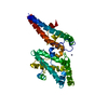 3vuvC  2wauS C: citing same article ( S: Starting model for refinement |
|---|---|
| Similar structure data |


-Links
 PDBj
PDBj- Assembly
| Deposited unit | 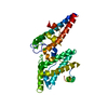
| ||||||||
|---|---|---|---|---|---|---|---|---|---|
| 1 |
| ||||||||
| Unit cell |
|
-Components
| #1: Protein | Red blood cell Mass: 35949.598 Da / Num. of mol.: 1 / Fragment: DBL domain, UNP residues 161-460 Source method: isolated from a genetically manipulated source Source: (gene. exp.) Plasmodium falciparum (malaria parasite P. falciparum)Strain: 3D7 / Gene: PF10_0355 / Production host:  Escherichia coli (E. coli) / References: UniProt: Q8IJ45 Escherichia coli (E. coli) / References: UniProt: Q8IJ45 | ||||
|---|---|---|---|---|---|
| #2: Chemical | Chloride  Mass: 35.453 Da / Num. of mol.: 2 / Source method: obtained synthetically / Formula: Cl Mass: 35.453 Da / Num. of mol.: 2 / Source method: obtained synthetically / Formula: Cl#3: Water | ChemComp-HOH / | Water Mass: 18.015 Da / Num. of mol.: 52 / Source method: isolated from a natural source / Formula: H2O Mass: 18.015 Da / Num. of mol.: 52 / Source method: isolated from a natural source / Formula: H2OSequence details | AN ARTEFACT OF THE TEV CLEAVAGE SITE INTRODUCED | |
-Experimental details
-Experiment
| Experiment | Method: X-RAY DIFFRACTION / Number of used crystals: 1 |
|---|
- Sample preparation
| Crystal | Density Matthews: 1.94 Å3/Da / Density % sol: 36.7 % Description: THE ENTRY CONTAINS FRIEDEL PAIRS IN F_PLUS/MINUS COLUMNS |
|---|---|
| Crystal grow | Temperature: 300 K / Method: vapor diffusion, sitting drop Details: 20% PEG3350, 0.2M sodium thiocyanate, VAPOR DIFFUSION, SITTING DROP, temperature 300K |
-Data collection
| Diffraction | Mean temperature: 100 K | |||||||||||||||||||||||||||||||||
|---|---|---|---|---|---|---|---|---|---|---|---|---|---|---|---|---|---|---|---|---|---|---|---|---|---|---|---|---|---|---|---|---|---|---|
| Diffraction source | Source: SYNCHROTRON / Site: Australian Synchrotron  / Beamline: MX2 / Wavelength: 1.0163 Å / Beamline: MX2 / Wavelength: 1.0163 Å | |||||||||||||||||||||||||||||||||
| Detector | Type: ADSC QUANTUM 315r / Detector: CCD / Date: Aug 25, 2010 | |||||||||||||||||||||||||||||||||
| Radiation | Monochromator: Si 111 CHANNEL / Protocol: SINGLE WAVELENGTH / Monochromatic (M) / Laue (L): M / Scattering type: x-ray | |||||||||||||||||||||||||||||||||
| Radiation wavelength | Wavelength: 1.0163 Å / Relative weight: 1 | |||||||||||||||||||||||||||||||||
| Reflection | Resolution: 2.09→50 Å / Num. all: 16232 / Num. obs: 16184 / % possible obs: 99.7 % / Observed criterion σ(F): -3 / Observed criterion σ(I): -3 | |||||||||||||||||||||||||||||||||
| Reflection shell |
|
- Processing
| Software |
| ||||||||||||||||||||||||||||||||||||||||||||||||||||||||||||||||||||||||||||||||||||||||||||||||||||||||||||||||||||||||||||||||||||||||||||||||||||||||||||||||||||||||||||||||||||||||||||||||||||||||
|---|---|---|---|---|---|---|---|---|---|---|---|---|---|---|---|---|---|---|---|---|---|---|---|---|---|---|---|---|---|---|---|---|---|---|---|---|---|---|---|---|---|---|---|---|---|---|---|---|---|---|---|---|---|---|---|---|---|---|---|---|---|---|---|---|---|---|---|---|---|---|---|---|---|---|---|---|---|---|---|---|---|---|---|---|---|---|---|---|---|---|---|---|---|---|---|---|---|---|---|---|---|---|---|---|---|---|---|---|---|---|---|---|---|---|---|---|---|---|---|---|---|---|---|---|---|---|---|---|---|---|---|---|---|---|---|---|---|---|---|---|---|---|---|---|---|---|---|---|---|---|---|---|---|---|---|---|---|---|---|---|---|---|---|---|---|---|---|---|---|---|---|---|---|---|---|---|---|---|---|---|---|---|---|---|---|---|---|---|---|---|---|---|---|---|---|---|---|---|---|---|---|
| Refinement | Method to determine structure: MOLECULAR REPLACEMENT Starting model: 2WAU Resolution: 2.093→37.583 Å / SU ML: 0.26 / σ(F): 1.91 / Phase error: 28.72 / Stereochemistry target values: ML Details: THE ENTRY CONTAINS FRIEDEL PAIRS IN F_PLUS/MINUS COLUMNS
| ||||||||||||||||||||||||||||||||||||||||||||||||||||||||||||||||||||||||||||||||||||||||||||||||||||||||||||||||||||||||||||||||||||||||||||||||||||||||||||||||||||||||||||||||||||||||||||||||||||||||
| Solvent computation | Shrinkage radii: 0.9 Å / VDW probe radii: 1.1 Å / Solvent model: FLAT BULK SOLVENT MODEL | ||||||||||||||||||||||||||||||||||||||||||||||||||||||||||||||||||||||||||||||||||||||||||||||||||||||||||||||||||||||||||||||||||||||||||||||||||||||||||||||||||||||||||||||||||||||||||||||||||||||||
| Refinement step | Cycle: LAST / Resolution: 2.093→37.583 Å
| ||||||||||||||||||||||||||||||||||||||||||||||||||||||||||||||||||||||||||||||||||||||||||||||||||||||||||||||||||||||||||||||||||||||||||||||||||||||||||||||||||||||||||||||||||||||||||||||||||||||||
| Refine LS restraints |
| ||||||||||||||||||||||||||||||||||||||||||||||||||||||||||||||||||||||||||||||||||||||||||||||||||||||||||||||||||||||||||||||||||||||||||||||||||||||||||||||||||||||||||||||||||||||||||||||||||||||||
| LS refinement shell | Refine-ID: X-RAY DIFFRACTION / Total num. of bins used: 10
| ||||||||||||||||||||||||||||||||||||||||||||||||||||||||||||||||||||||||||||||||||||||||||||||||||||||||||||||||||||||||||||||||||||||||||||||||||||||||||||||||||||||||||||||||||||||||||||||||||||||||
| Refinement TLS params. | Method: refined / Refine-ID: X-RAY DIFFRACTION
| ||||||||||||||||||||||||||||||||||||||||||||||||||||||||||||||||||||||||||||||||||||||||||||||||||||||||||||||||||||||||||||||||||||||||||||||||||||||||||||||||||||||||||||||||||||||||||||||||||||||||
| Refinement TLS group |
|
