 Movie
Movie Controller


+ Open data

- Basic information
| Entry | Database: PDB / ID: 3vuc | ||||||
|---|---|---|---|---|---|---|---|
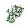
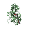
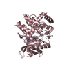
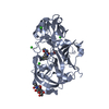
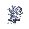
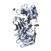
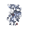
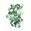
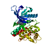
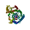
| Title | Human renin in complex with compound 5 | ||||||
 Components Components | Renin | ||||||
 Keywords Keywords | HYDROLASE/HYDROLASE INHIBITOR / Aspartyl protease / RAS / hypertension / inhibitor / HYDROLASE-HYDROLASE INHIBITOR complex | ||||||
| Function / homology |  Function and homology informationrenin / juxtaglomerular apparatus development / mesonephros development / response to cGMP / renin-angiotensin regulation of aldosterone production / drinking behavior / regulation of MAPK cascade / response to immobilization stress / angiotensin maturation / amyloid-beta metabolic process ...renin / juxtaglomerular apparatus development / mesonephros development / response to cGMP / renin-angiotensin regulation of aldosterone production / drinking behavior / regulation of MAPK cascade / response to immobilization stress / angiotensin maturation / amyloid-beta metabolic process / Metabolism of Angiotensinogen to Angiotensins / cell maturation / response to cAMP / insulin-like growth factor receptor binding / hormone-mediated signaling pathway / kidney development / regulation of blood pressure / male gonad development / cellular response to xenobiotic stimulus / apical part of cell / peptidase activity / response to lipopolysaccharide / aspartic-type endopeptidase activity / signaling receptor binding / proteolysis / extracellular space / extracellular region / plasma membrane Function and homology informationrenin / juxtaglomerular apparatus development / mesonephros development / response to cGMP / renin-angiotensin regulation of aldosterone production / drinking behavior / regulation of MAPK cascade / response to immobilization stress / angiotensin maturation / amyloid-beta metabolic process ...renin / juxtaglomerular apparatus development / mesonephros development / response to cGMP / renin-angiotensin regulation of aldosterone production / drinking behavior / regulation of MAPK cascade / response to immobilization stress / angiotensin maturation / amyloid-beta metabolic process / Metabolism of Angiotensinogen to Angiotensins / cell maturation / response to cAMP / insulin-like growth factor receptor binding / hormone-mediated signaling pathway / kidney development / regulation of blood pressure / male gonad development / cellular response to xenobiotic stimulus / apical part of cell / peptidase activity / response to lipopolysaccharide / aspartic-type endopeptidase activity / signaling receptor binding / proteolysis / extracellular space / extracellular region / plasma membraneSimilarity search - Function | ||||||
| Biological species |  Homo sapiens (human) Homo sapiens (human) | ||||||
| Method | X-RAY DIFFRACTION / SYNCHROTRON / MOLECULAR REPLACEMENT / Resolution: 2.6 Å | ||||||
 Authors Authors | Takahashi, M. / Matsui, Y. / Hanzawa, H. | ||||||
 Citation Citation | Journal: Acs Med.Chem.Lett. / Year: 2012 Title: Discovery of DS-8108b, a Novel Orally Bioavailable Renin Inhibitor. Authors: Nakamura, Y. / Fujimoto, T. / Ogawa, Y. / Sugita, C. / Miyazaki, S. / Tamaki, K. / Takahashi, M. / Matsui, Y. / Nagayama, T. / Manabe, K. / Mizuno, M. / Masubuchi, N. / Chiba, K. / Nishi, T. | ||||||
| History |
|
- Structure visualization
| Structure viewer | Molecule: MolmilJmol/JSmol |
|---|
- Downloads & links
-Download
| PDBx/mmCIF format | 3vuc.cif.gz | 274.8 KB | Display | PDBx/mmCIF format |
|---|---|---|---|---|
| PDB format | pdb3vuc.ent.gz | 224.6 KB | Display | PDB format |
| PDBx/mmJSON format | 3vuc.json.gz | Tree view | PDBx/mmJSON format | |
| Others |  Other downloads Other downloads |
-Validation report
| Arichive directory | https://data.pdbj.org/pub/pdb/validation_reports/vu/3vucftp://data.pdbj.org/pub/pdb/validation_reports/vu/3vuc | HTTPS FTP |
|---|
-Related structure data
| Related structure data |  3vsxS S: Starting model for refinement |
|---|---|
| Similar structure data |


-Links
 PDBj
PDBj


- Assembly
| Deposited unit | 
| ||||||||
|---|---|---|---|---|---|---|---|---|---|
| 1 | 
| ||||||||
| 2 | 
| ||||||||
| 3 | 
| ||||||||
| Unit cell |
|
-Components
| #1: Protein | / Angiotensinogenase Mass: 37267.008 Da / Num. of mol.: 2 Source method: isolated from a genetically manipulated source Source: (gene. exp.) Homo sapiens (human) / Gene: REN / Plasmid: pcDNA3.1 / Cell line (production host): 293F / Production host: Homo sapiens (human) / References: UniProt: P00797, renin#2: Sugar | N-Acetylglucosamine  Type: D-saccharide, beta linking / Mass: 221.208 Da / Num. of mol.: 2 Type: D-saccharide, beta linking / Mass: 221.208 Da / Num. of mol.: 2Source method: isolated from a genetically manipulated source Formula: C8H15NO6 #3: Chemical |   Mass: 561.156 Da / Num. of mol.: 2 / Source method: obtained synthetically / Formula: C30H45ClN4O4 Mass: 561.156 Da / Num. of mol.: 2 / Source method: obtained synthetically / Formula: C30H45ClN4O4#4: Water | ChemComp-HOH / | Water Mass: 18.015 Da / Num. of mol.: 212 / Source method: isolated from a natural source / Formula: H2O Mass: 18.015 Da / Num. of mol.: 212 / Source method: isolated from a natural source / Formula: H2O |
|---|
-Experimental details
-Experiment
| Experiment | Method: X-RAY DIFFRACTION / Number of used crystals: 1 |
|---|
- Sample preparation
| Crystal | Density Matthews: 3.15 Å3/Da / Density % sol: 61 % / Mosaicity: 0.387 ° |
|---|---|
| Crystal grow | Temperature: 295 K / Method: vapor diffusion, hanging drop / pH: 3 Details: 5-12% PEG 3350, 0.6M NaCl, 0.1M citrate pH3.0-4.5, vapor diffusion, hanging drop, temperature 295K |
-Data collection
| Diffraction | Mean temperature: 100 K | |||||||||||||||||||||||||||||||||||||||||||||||||||||||||||||||||||||||||||||
|---|---|---|---|---|---|---|---|---|---|---|---|---|---|---|---|---|---|---|---|---|---|---|---|---|---|---|---|---|---|---|---|---|---|---|---|---|---|---|---|---|---|---|---|---|---|---|---|---|---|---|---|---|---|---|---|---|---|---|---|---|---|---|---|---|---|---|---|---|---|---|---|---|---|---|---|---|---|---|
| Diffraction source | Source: SYNCHROTRON / Site: Photon Factory  / Beamline: BL-5A / Wavelength: 1 Å / Beamline: BL-5A / Wavelength: 1 Å | |||||||||||||||||||||||||||||||||||||||||||||||||||||||||||||||||||||||||||||
| Detector | Type: ADSC QUANTUM 210r / Detector: CCD / Date: Oct 5, 2010 | |||||||||||||||||||||||||||||||||||||||||||||||||||||||||||||||||||||||||||||
| Radiation | Protocol: SINGLE WAVELENGTH / Monochromatic (M) / Laue (L): M / Scattering type: x-ray | |||||||||||||||||||||||||||||||||||||||||||||||||||||||||||||||||||||||||||||
| Radiation wavelength | Wavelength: 1 Å / Relative weight: 1 | |||||||||||||||||||||||||||||||||||||||||||||||||||||||||||||||||||||||||||||
| Reflection | Resolution: 2.6→50 Å / Num. obs: 29254 / % possible obs: 99.9 % / Redundancy: 11 % / Rmerge(I) obs: 0.064 / Χ2: 1.171 / Net I/σ(I): 15.5 | |||||||||||||||||||||||||||||||||||||||||||||||||||||||||||||||||||||||||||||
| Reflection shell |
|
- Processing
| Software |
| |||||||||||||||||||||||||||||||||||||||||||||||||||||||||||||||||||||||||||||||||||||||||||||||||||||||||||||||||||||||||||||
|---|---|---|---|---|---|---|---|---|---|---|---|---|---|---|---|---|---|---|---|---|---|---|---|---|---|---|---|---|---|---|---|---|---|---|---|---|---|---|---|---|---|---|---|---|---|---|---|---|---|---|---|---|---|---|---|---|---|---|---|---|---|---|---|---|---|---|---|---|---|---|---|---|---|---|---|---|---|---|---|---|---|---|---|---|---|---|---|---|---|---|---|---|---|---|---|---|---|---|---|---|---|---|---|---|---|---|---|---|---|---|---|---|---|---|---|---|---|---|---|---|---|---|---|---|---|---|
| Refinement | Method to determine structure: MOLECULAR REPLACEMENT Starting model: PDB ENTRY 3VSX Resolution: 2.6→20 Å / Cor.coef. Fo:Fc: 0.947 / Cor.coef. Fo:Fc free: 0.907 / Occupancy max: 1 / Occupancy min: 1 / SU B: 20.6 / SU ML: 0.207 / Cross valid method: THROUGHOUT / σ(F): 0 / ESU R: 0.604 / ESU R Free: 0.303 / Stereochemistry target values: MAXIMUM LIKELIHOOD
| |||||||||||||||||||||||||||||||||||||||||||||||||||||||||||||||||||||||||||||||||||||||||||||||||||||||||||||||||||||||||||||
| Solvent computation | Ion probe radii: 0.8 Å / Shrinkage radii: 0.8 Å / VDW probe radii: 1.4 Å / Solvent model: BABINET MODEL WITH MASK | |||||||||||||||||||||||||||||||||||||||||||||||||||||||||||||||||||||||||||||||||||||||||||||||||||||||||||||||||||||||||||||
| Displacement parameters | Biso max: 111.4 Å2 / Biso mean: 49.0698 Å2 / Biso min: 10.5 Å2 | |||||||||||||||||||||||||||||||||||||||||||||||||||||||||||||||||||||||||||||||||||||||||||||||||||||||||||||||||||||||||||||
| Refinement step | Cycle: LAST / Resolution: 2.6→20 Å
| |||||||||||||||||||||||||||||||||||||||||||||||||||||||||||||||||||||||||||||||||||||||||||||||||||||||||||||||||||||||||||||
| Refine LS restraints |
| |||||||||||||||||||||||||||||||||||||||||||||||||||||||||||||||||||||||||||||||||||||||||||||||||||||||||||||||||||||||||||||
| LS refinement shell | Resolution: 2.6→2.666 Å / Total num. of bins used: 20
| |||||||||||||||||||||||||||||||||||||||||||||||||||||||||||||||||||||||||||||||||||||||||||||||||||||||||||||||||||||||||||||
| Refinement TLS params. | Method: refined / Refine-ID: X-RAY DIFFRACTION
| |||||||||||||||||||||||||||||||||||||||||||||||||||||||||||||||||||||||||||||||||||||||||||||||||||||||||||||||||||||||||||||
| Refinement TLS group |
|
