 Movie
Movie Controller

[English] 日本語
 Yorodumi
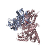
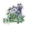
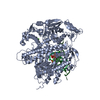
- PDB-3vpb: ArgX from Sulfolobus tokodaii complexed with LysW/Glu/ADP/Mg/Zn/S... -
+ Open data

- Basic information
| Entry | Database: PDB / ID: 3vpb | ||||||
|---|---|---|---|---|---|---|---|
| Title | ArgX from Sulfolobus tokodaii complexed with LysW/Glu/ADP/Mg/Zn/Sulfate | ||||||
 Components Components |
| ||||||
 Keywords Keywords |  LIGASE / ATP-dependent amine/thiol ligase family / ATP-dependent amine/thiol ligase / LysW / Enzyme-Carrier protein complex LIGASE / ATP-dependent amine/thiol ligase family / ATP-dependent amine/thiol ligase / LysW / Enzyme-Carrier protein complex | ||||||
| Function / homology |  Function and homology information Function and homology informationlysine biosynthetic process / Ligases; Forming carbon-nitrogen bonds; Acid-amino-acid ligases (peptide synthases) / lysine biosynthetic process via aminoadipic acid / arginine biosynthetic process / ligase activity / protein modification process / ATP binding / identical protein binding / metal ion bindingSimilarity search - Function | ||||||
| Biological species |   Sulfolobus tokodaii (archaea) Sulfolobus tokodaii (archaea) | ||||||
| Method | X-RAY DIFFRACTION / SYNCHROTRON / MOLECULAR REPLACEMENT / Resolution: 1.8 Å | ||||||
 Authors Authors | Tomita, T. / Ouchi, T. / Kuzuyama, T. / Nishiyama, M. | ||||||
 Citation Citation | Journal: Nat.Chem.Biol. / Year: 2013 Title: Lysine and arginine biosyntheses mediated by a common carrier protein in Sulfolobus Authors: Ouchi, T. / Tomita, T. / Horie, A. / Yoshida, A. / Takahashi, K. / Nishida, H. / Lassak, K. / Taka, H. / Mineki, R. / Fujimura, T. / Kosono, S. / Nishiyama, C. / Masui, R. / Kuramitsu, S. / ...Authors: Ouchi, T. / Tomita, T. / Horie, A. / Yoshida, A. / Takahashi, K. / Nishida, H. / Lassak, K. / Taka, H. / Mineki, R. / Fujimura, T. / Kosono, S. / Nishiyama, C. / Masui, R. / Kuramitsu, S. / Albers, S.V. / Kuzuyama, T. / Nishiyama, M. | ||||||
| History |
|
- Structure visualization
| Structure viewer | Molecule: MolmilJmol/JSmol |
|---|
- Downloads & links
-Download
| PDBx/mmCIF format | 3vpb.cif.gz | 280.5 KB | Display | PDBx/mmCIF format |
|---|---|---|---|---|
| PDB format | pdb3vpb.ent.gz | 223.6 KB | Display | PDB format |
| PDBx/mmJSON format | 3vpb.json.gz | Tree view | PDBx/mmJSON format | |
| Others |  Other downloads Other downloads |
-Validation report
| Arichive directory | https://data.pdbj.org/pub/pdb/validation_reports/vp/3vpbftp://data.pdbj.org/pub/pdb/validation_reports/vp/3vpb | HTTPS FTP |
|---|
-Related structure data
| Related structure data | 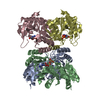 3vpcC 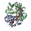 3vpdC 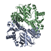 1uc8S C: citing same article ( S: Starting model for refinement |
|---|---|
| Similar structure data |


-Links
 PDBj
PDBj


- Assembly
| Deposited unit | 
| ||||||||
|---|---|---|---|---|---|---|---|---|---|
| 1 |
| ||||||||
| Unit cell |
|
-Components
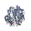
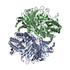
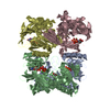
-Protein , 2 types, 6 molecules ABCDEF
| #1: Protein | Mass: 31572.408 Da / Num. of mol.: 4 Source method: isolated from a genetically manipulated source Source: (gene. exp.) Sulfolobus tokodaii (archaea) / Strain: DSM 16993, JCM 10545, NBRC 100140, 7 / Gene: argE, STK_15050 / Plasmid: pET-26b(+), pET-StArgX / Production host:  Escherichia coli (E. coli) / Strain (production host): BL21CodonPlus(DE3) / References: UniProt: Q970U6, acetylornithine deacetylase Escherichia coli (E. coli) / Strain (production host): BL21CodonPlus(DE3) / References: UniProt: Q970U6, acetylornithine deacetylase#2: Protein | Mass: 6108.802 Da / Num. of mol.: 2 Source method: isolated from a genetically manipulated source Source: (gene. exp.) Sulfolobus tokodaii (archaea) / Strain: DSM 16993, JCM 10545, NBRC 100140, 7 / Gene: lysW, STK_01925, STS023 / Plasmid: pET-26b(+), pET-StLysW / Production host: Escherichia coli (E. coli) / Strain (production host): BL21CodonPlus(DE3) / References: UniProt: Q976J8 |
|---|
-Non-polymers , 6 types, 1062 molecules 


| #3: Chemical | ChemComp-ADP / Adenosine diphosphate Mass: 427.201 Da / Num. of mol.: 4 / Source method: obtained synthetically / Formula: C10H15N5O10P2 / Comment: ADP, energy-carrying molecule*YM Mass: 427.201 Da / Num. of mol.: 4 / Source method: obtained synthetically / Formula: C10H15N5O10P2 / Comment: ADP, energy-carrying molecule*YM#4: Chemical | ChemComp-GLU / Glutamic acid Type: L-peptide linking / Mass: 147.129 Da / Num. of mol.: 4 / Source method: obtained synthetically / Formula: C5H9NO4 Type: L-peptide linking / Mass: 147.129 Da / Num. of mol.: 4 / Source method: obtained synthetically / Formula: C5H9NO4#5: Chemical | ChemComp-MG /  Mass: 24.305 Da / Num. of mol.: 4 / Source method: obtained synthetically / Formula: Mg Mass: 24.305 Da / Num. of mol.: 4 / Source method: obtained synthetically / Formula: Mg#6: Chemical | ChemComp-ZN /  Mass: 65.409 Da / Num. of mol.: 4 / Source method: obtained synthetically / Formula: Zn Mass: 65.409 Da / Num. of mol.: 4 / Source method: obtained synthetically / Formula: Zn#7: Chemical | ChemComp-SO4 / Sulfate Mass: 96.063 Da / Num. of mol.: 6 / Source method: obtained synthetically / Formula: SO4 Mass: 96.063 Da / Num. of mol.: 6 / Source method: obtained synthetically / Formula: SO4#8: Water | ChemComp-HOH / | WaterMass: 18.015 Da / Num. of mol.: 1040 / Source method: isolated from a natural source / Formula: H2O |
|---|
-Experimental details
-Experiment
| Experiment | Method: X-RAY DIFFRACTION / Number of used crystals: 1 |
|---|
- Sample preparation
| Crystal | Density Matthews: 2.23 Å3/Da / Density % sol: 44.78 % |
|---|---|
| Crystal grow | Temperature: 293 K / Method: vapor diffusion, hanging drop / pH: 6 Details: 13% PEG-MME 5000, 0.1M Ammonium sulfate, 0.1M MES-NaOH (pH6.0), VAPOR DIFFUSION, HANGING DROP, temperature 293K |
-Data collection
| Diffraction | Mean temperature: 95 K |
|---|---|
| Diffraction source | Source: SYNCHROTRON / Site: Photon Factory  / Beamline: AR-NW12A / Wavelength: 1 Å / Beamline: AR-NW12A / Wavelength: 1 Å |
| Detector | Type: ADSC QUANTUM 210r / Detector: CCD / Date: Oct 9, 2010 |
| Radiation | Monochromator: MIRROR / Protocol: SINGLE WAVELENGTH / Monochromatic (M) / Laue (L): M / Scattering type: x-ray |
| Radiation wavelength | Wavelength: 1 Å / Relative weight: 1 |
| Reflection | Resolution: 1.8→50 Å / Num. all: 106366 / Num. obs: 106366 / % possible obs: 99.7 % / Observed criterion σ(F): 2 / Observed criterion σ(I): 2 |
| Reflection shell | Resolution: 1.8→1.85 Å / % possible all: 99.1 |
- Processing
| Software |
| |||||||||||||||||||||||||||||||||||||||||||||||||||||||||||||||||
|---|---|---|---|---|---|---|---|---|---|---|---|---|---|---|---|---|---|---|---|---|---|---|---|---|---|---|---|---|---|---|---|---|---|---|---|---|---|---|---|---|---|---|---|---|---|---|---|---|---|---|---|---|---|---|---|---|---|---|---|---|---|---|---|---|---|---|
| Refinement | Method to determine structure: MOLECULAR REPLACEMENT Starting model: 1UC8 Resolution: 1.8→28.91 Å / Cor.coef. Fo:Fc: 0.957 / Cor.coef. Fo:Fc free: 0.933 / SU B: 2.958 / SU ML: 0.094 / Cross valid method: THROUGHOUT / σ(F): 2 / ESU R: 0.144 / ESU R Free: 0.139 / Stereochemistry target values: MAXIMUM LIKELIHOOD / Details: HYDROGENS HAVE BEEN ADDED IN THE RIDING POSITIONS
| |||||||||||||||||||||||||||||||||||||||||||||||||||||||||||||||||
| Solvent computation | Ion probe radii: 0.8 Å / Shrinkage radii: 0.8 Å / VDW probe radii: 1.4 Å / Solvent model: MASK | |||||||||||||||||||||||||||||||||||||||||||||||||||||||||||||||||
| Displacement parameters | Biso mean: 24.167 Å2
| |||||||||||||||||||||||||||||||||||||||||||||||||||||||||||||||||
| Refinement step | Cycle: LAST / Resolution: 1.8→28.91 Å
| |||||||||||||||||||||||||||||||||||||||||||||||||||||||||||||||||
| Refine LS restraints |
| |||||||||||||||||||||||||||||||||||||||||||||||||||||||||||||||||
| LS refinement shell | Resolution: 1.798→1.845 Å / Total num. of bins used: 20
|
