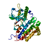
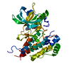
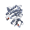
Entry Database : PDB / ID : 3vhkTitle Crystal structure of the VEGFR2 kinase domain in complex with a back pocket binder Vascular endothelial growth factor receptor 2 Keywords / / / Function / homology Function Domain/homology Component
/ / / / / / / / / / / / / / / / / / / / / / / / / / / / / / / / / / / / / / / / / / / / / / / / / / / / / / / / / / / / / / / / / / / / / / / / / / / / / / / / / / / / / / / / / / / / / / / / / / / / / / / / / / / / / / / / / / / / / / / / / / / / / / / / / / / / / / / / / / / / / / / / / / / / Biological species Homo sapiens (human)Method / / / / Resolution : 2.49 Å Authors Iwata, H. / Oki, H. / Okada, K. / Takagi, T. / Tawada, M. / Miyazaki, Y. / Imamura, S. / Hori, A. / Hixon, M.S. / Kimura, H. / Miki, H. Journal : ACS MED.CHEM.LETT. / Year : 2012Title : A Back-to-Front Fragment-Based Drug Design Search Strategy Targeting the DFG-Out Pocket of Protein Tyrosine Kinases.Authors : Iwata, H. / Oki, H. / Okada, K. / Takagi, T. / Tawada, M. / Miyazaki, Y. / Imamura, S. / Hori, A. / Lawson, J.D. / Hixon, M.S. / Kimura, H. / Miki, H. History Deposition Aug 25, 2011 Deposition site / Processing site Revision 1.0 Sep 5, 2012 Provider / Type Revision 1.1 Apr 22, 2015 Group Revision 1.2 Mar 20, 2024 Group / Database references / Derived calculationsCategory chem_comp_atom / chem_comp_bond ... chem_comp_atom / chem_comp_bond / database_2 / struct_ref_seq_dif / struct_site Item _database_2.pdbx_DOI / _database_2.pdbx_database_accession ... _database_2.pdbx_DOI / _database_2.pdbx_database_accession / _struct_ref_seq_dif.details / _struct_site.pdbx_auth_asym_id / _struct_site.pdbx_auth_comp_id / _struct_site.pdbx_auth_seq_id
Show all Show less
 Movie
Movie Controller

 Yorodumi
 Open data
 Basic information
 Components
 VEGF receptor
  Keywords
 Function and homology information
 Authors
 Citation
 Structure visualization
 Downloads & links
 Other downloads


 PDBj
PDBj


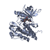
 Assembly
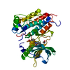


 Mass: 295.332 Da / Num. of mol.: 1 / Source method: obtained synthetically / Formula: C18H17NO3
 Mass: 62.068 Da / Num. of mol.: 1 / Source method: obtained synthetically / Formula: C2H6O2
 Mass: 18.015 Da / Num. of mol.: 42 / Source method: isolated from a natural source / Formula: H2O
 Sample preparation
 / Beamline: BL32B2 / Wavelength: 1 Å
 Processing