 Movie
Movie Controller

[English] 日本語
 Yorodumi
- PDB-3v3y: Photosynthetic Reaction Center From Rhodobacter Sphaeroides strain RV -
+ Open data

- Basic information
| Entry | Database: PDB / ID: 3v3y | ||||||
|---|---|---|---|---|---|---|---|
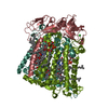
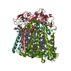
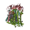
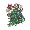
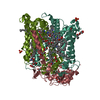
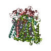
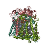
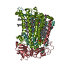
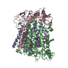
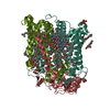
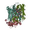
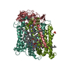
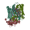
| Title | Photosynthetic Reaction Center From Rhodobacter Sphaeroides strain RV | ||||||
 Components Components | (Reaction center protein ... Photosynthetic reaction centre) x 3 Photosynthetic reaction centre) x 3 | ||||||
 Keywords Keywords | ELECTRON TRANSPORT / Photosynthetic reaction center / Primary electron donor / membrane | ||||||
| Function / homology |  Function and homology information Function and homology informationplasma membrane-derived chromatophore membrane / plasma membrane light-harvesting complex / bacteriochlorophyll binding / photosynthesis, light reaction / electron transporter, transferring electrons within the cyclic electron transport pathway of photosynthesis activity / photosynthetic electron transport in photosystem II / metal ion bindingSimilarity search - Function | ||||||
| Biological species |  Rhodobacter sphaeroides (bacteria) Rhodobacter sphaeroides (bacteria) | ||||||
| Method | X-RAY DIFFRACTION / MOLECULAR REPLACEMENT / Resolution: 2.8 Å | ||||||
 Authors Authors | Gabdulkhakov, A.G. / Fufina, T.Y. / Vasilieva, L.G. / Shuvalov, V.A. | ||||||
 Citation Citation | Journal: Biochim.Biophys.Acta / Year: 2012 Title: The site-directed mutation I(L177)H in Rhodobacter sphaeroides reaction center affects coordination of P(A) and B(B) bacteriochlorophylls. Authors: Vasilieva, L.G. / Fufina, T.Y. / Gabdulkhakov, A.G. / Leonova, M.M. / Khatypov, R.A. / Shuvalov, V.A. | ||||||
| History |
|
- Structure visualization
| Structure viewer | Molecule: MolmilJmol/JSmol |
|---|
- Downloads & links
-Download
| PDBx/mmCIF format | 3v3y.cif.gz | 201.6 KB | Display | PDBx/mmCIF format |
|---|---|---|---|---|
| PDB format | pdb3v3y.ent.gz | 156.8 KB | Display | PDB format |
| PDBx/mmJSON format | 3v3y.json.gz | Tree view | PDBx/mmJSON format | |
| Others |  Other downloads Other downloads |
-Validation report
| Arichive directory | https://data.pdbj.org/pub/pdb/validation_reports/v3/3v3yftp://data.pdbj.org/pub/pdb/validation_reports/v3/3v3y | HTTPS FTP |
|---|
-Related structure data
| Related structure data |  3v3zC  1e6dS S: Starting model for refinement C: citing same article ( |
|---|---|
| Similar structure data |


-Links
 PDBj
PDBj


- Assembly
| Deposited unit | 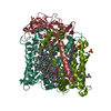
| ||||||||
|---|---|---|---|---|---|---|---|---|---|
| 1 |
| ||||||||
| Unit cell |
| ||||||||
| Details | heterotrimer |
-Components
-Reaction center protein ... , 3 types, 3 molecules HLM
| #1: Protein | Photosynthetic reaction centre / Photosynthetic reaction center H subunit Mass: 26170.078 Da / Num. of mol.: 1 Source method: isolated from a genetically manipulated source Source: (gene. exp.) Rhodobacter sphaeroides (bacteria) / Gene: puhA / Plasmid: pREH-D2 L177 / Production host: Rhodobacter sphaeroides (bacteria) / Strain (production host): DD13 / References: UniProt: P0C0Y7 |
|---|---|
| #2: Protein | Photosynthetic reaction centre / Photosynthetic reaction center L subunit Mass: 31360.416 Da / Num. of mol.: 1 Source method: isolated from a genetically manipulated source Source: (gene. exp.) Rhodobacter sphaeroides (bacteria) / Gene: pufL / Plasmid: pREH-D2 L177 / Production host: Rhodobacter sphaeroides (bacteria) / Strain (production host): DD13 / References: UniProt: P0C0Y8 |
| #3: Protein | Photosynthetic reaction centre / Photosynthetic reaction center M subunit Mass: 33885.922 Da / Num. of mol.: 1 Source method: isolated from a genetically manipulated source Source: (gene. exp.) Rhodobacter sphaeroides (bacteria) / Gene: pufM / Plasmid: pREH-D2 L177 / Production host: Rhodobacter sphaeroides (bacteria) / Strain (production host): DD13 / References: UniProt: P0C0Y9 |
-Non-polymers , 10 types, 117 molecules 


| #4: Chemical | ChemComp-LDA / Lauryldimethylamine oxide Mass: 229.402 Da / Num. of mol.: 8 / Source method: obtained synthetically / Formula: C14H31NO / Comment: LDAO, detergent*YM Mass: 229.402 Da / Num. of mol.: 8 / Source method: obtained synthetically / Formula: C14H31NO / Comment: LDAO, detergent*YM#5: Chemical | ChemComp-BCL / Bacteriochlorophyll Mass: 911.504 Da / Num. of mol.: 4 / Source method: obtained synthetically / Formula: C55H74MgN4O6 Mass: 911.504 Da / Num. of mol.: 4 / Source method: obtained synthetically / Formula: C55H74MgN4O6#6: Chemical | Pheophytin Mass: 889.215 Da / Num. of mol.: 2 / Source method: obtained synthetically / Formula: C55H76N4O6 Mass: 889.215 Da / Num. of mol.: 2 / Source method: obtained synthetically / Formula: C55H76N4O6#7: Chemical | Coenzyme Q10 Mass: 863.343 Da / Num. of mol.: 2 / Source method: obtained synthetically / Formula: C59H90O4 Mass: 863.343 Da / Num. of mol.: 2 / Source method: obtained synthetically / Formula: C59H90O4#8: Chemical | 1,4-Dioxane Mass: 88.105 Da / Num. of mol.: 3 / Source method: obtained synthetically / Formula: C4H8O2 Mass: 88.105 Da / Num. of mol.: 3 / Source method: obtained synthetically / Formula: C4H8O2#9: Chemical | ChemComp-FE / | Iron Mass: 55.845 Da / Num. of mol.: 1 / Source method: obtained synthetically / Formula: Fe Mass: 55.845 Da / Num. of mol.: 1 / Source method: obtained synthetically / Formula: Fe#10: Chemical | ChemComp-SPN / |  Mass: 594.993 Da / Num. of mol.: 1 / Source method: obtained synthetically / Formula: C41H70O2 Mass: 594.993 Da / Num. of mol.: 1 / Source method: obtained synthetically / Formula: C41H70O2#11: Chemical | Phosphate Mass: 94.971 Da / Num. of mol.: 3 / Source method: obtained synthetically / Formula: PO4 Mass: 94.971 Da / Num. of mol.: 3 / Source method: obtained synthetically / Formula: PO4#12: Chemical | ChemComp-CL / | Chloride Mass: 35.453 Da / Num. of mol.: 1 / Source method: obtained synthetically / Formula: Cl Mass: 35.453 Da / Num. of mol.: 1 / Source method: obtained synthetically / Formula: Cl#13: Water | ChemComp-HOH / | WaterMass: 18.015 Da / Num. of mol.: 92 / Source method: isolated from a natural source / Formula: H2O |
|---|
-Details
| Sequence details | AUTHORS STATE THAT THE DISCREPANC |
|---|
-Experimental details
-Experiment
| Experiment | Method: X-RAY DIFFRACTION / Number of used crystals: 1 |
|---|
- Sample preparation
| Crystal | Density Matthews: 5.72 Å3/Da / Density % sol: 78.51 % |
|---|---|
| Crystal grow | Temperature: 289 K / Method: vapor diffusion, hanging drop / pH: 7.4 Details: 3.5% 1,2,3 -heptanetriol, 2% dioxane, 0.1% LDAO, 1M potassium phosphate , pH 7.4, VAPOR DIFFUSION, HANGING DROP, temperature 289K |
-Data collection
| Diffraction | Mean temperature: 110 K |
|---|---|
| Diffraction source | Source: ROTATING ANODE / Type: BRUKER AXS MICROSTAR / Wavelength: 1.54179 Å |
| Detector | Type: Bruker Platinum 135 / Detector: CCD / Date: Nov 11, 2011 |
| Radiation | Monochromator: Montel 200 / Protocol: SINGLE WAVELENGTH / Monochromatic (M) / Laue (L): M / Scattering type: x-ray |
| Radiation wavelength | Wavelength: 1.54179 Å / Relative weight: 1 |
| Reflection | Resolution: 2.8→26.6 Å / Num. all: 52181 / Num. obs: 51787 / % possible obs: 99.2 % / Observed criterion σ(F): 0 / Observed criterion σ(I): 0 / Redundancy: 3.75 % / Rmerge(I) obs: 0.185 / Net I/σ(I): 6.11 |
| Reflection shell | Resolution: 2.8→2.9 Å / Redundancy: 3.91 % / Rmerge(I) obs: 0.626 / Mean I/σ(I) obs: 1.7 / % possible all: 99.5 |
- Processing
| Software |
| ||||||||||||||||||||||||||||||||||||||||||||||||||||||||||||||||||||||||||||||||||||||||||||||||||||||||||||||||||||||||||||||||||||||||||||
|---|---|---|---|---|---|---|---|---|---|---|---|---|---|---|---|---|---|---|---|---|---|---|---|---|---|---|---|---|---|---|---|---|---|---|---|---|---|---|---|---|---|---|---|---|---|---|---|---|---|---|---|---|---|---|---|---|---|---|---|---|---|---|---|---|---|---|---|---|---|---|---|---|---|---|---|---|---|---|---|---|---|---|---|---|---|---|---|---|---|---|---|---|---|---|---|---|---|---|---|---|---|---|---|---|---|---|---|---|---|---|---|---|---|---|---|---|---|---|---|---|---|---|---|---|---|---|---|---|---|---|---|---|---|---|---|---|---|---|---|---|---|
| Refinement | Method to determine structure: MOLECULAR REPLACEMENT Starting model: 1E6D Resolution: 2.8→19.941 Å / SU ML: 0.9 / σ(F): 0 / Phase error: 28.91 / Stereochemistry target values: ML
| ||||||||||||||||||||||||||||||||||||||||||||||||||||||||||||||||||||||||||||||||||||||||||||||||||||||||||||||||||||||||||||||||||||||||||||
| Solvent computation | Shrinkage radii: 0.47 Å / VDW probe radii: 0.8 Å / Solvent model: FLAT BULK SOLVENT MODEL / Bsol: 11.091 Å2 / ksol: 0.274 e/Å3 | ||||||||||||||||||||||||||||||||||||||||||||||||||||||||||||||||||||||||||||||||||||||||||||||||||||||||||||||||||||||||||||||||||||||||||||
| Displacement parameters |
| ||||||||||||||||||||||||||||||||||||||||||||||||||||||||||||||||||||||||||||||||||||||||||||||||||||||||||||||||||||||||||||||||||||||||||||
| Refinement step | Cycle: LAST / Resolution: 2.8→19.941 Å
| ||||||||||||||||||||||||||||||||||||||||||||||||||||||||||||||||||||||||||||||||||||||||||||||||||||||||||||||||||||||||||||||||||||||||||||
| Refine LS restraints |
| ||||||||||||||||||||||||||||||||||||||||||||||||||||||||||||||||||||||||||||||||||||||||||||||||||||||||||||||||||||||||||||||||||||||||||||
| LS refinement shell |
|
