- PDB-1rgn: Structure of the reaction centre from Rhodobacter sphaeroides car... -
+
Open data
ID or keywords:
Loading...
-
Basic information
Entry
Database: PDB / ID: 1rgn
Title
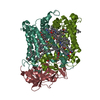
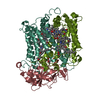
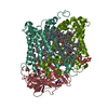
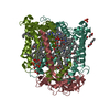
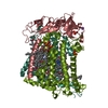
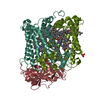
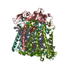
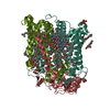
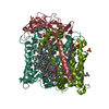
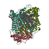
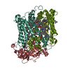
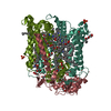
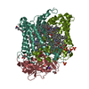
Structure of the reaction centre from Rhodobacter sphaeroides carotenoidless strain R-26.1 reconstituted with spheroidene
Components
(Reaction center protein ...Photosynthetic reaction centre) x 3
Keywords
PHOTOSYNTHESIS / PHOTOSYNTHETIC REACTION CENTER / RECONSTITUTED CAROTENOID / CAROTENOID BINDING SITE / MEMBRANE PROTEIN
Function / homology
Function and homology information
: / plasma membrane-derived chromatophore membrane / plasma membrane light-harvesting complex / bacteriochlorophyll binding / photosynthesis, light reaction / electron transporter, transferring electrons within the cyclic electron transport pathway of photosynthesis activity / photosynthetic electron transport in photosystem II / membrane => GO:0016020 / metal ion binding Similarity search - Function
Photosynthetic Reaction Center, subunit M; domain 1 / Photosystem II protein D1-like / Photosynthetic Reaction Center; Chain H, domain 2 / Photosynthetic Reaction Center, subunit H, domain 2 / Photosynthetic Reaction Center; Chain H, domain 1 / Photosynthetic reaction centre, H subunit, N-terminal domain / Photosynthetic reaction centre, H subunit / Bacterial photosynthetic reaction centre, H-chain, C-terminal / Photosynthetic reaction centre, M subunit / Photosynthetic reaction centre, H subunit, N-terminal ...Photosynthetic Reaction Center, subunit M; domain 1 / Photosystem II protein D1-like / Photosynthetic Reaction Center; Chain H, domain 2 / Photosynthetic Reaction Center, subunit H, domain 2 / Photosynthetic Reaction Center; Chain H, domain 1 / Photosynthetic reaction centre, H subunit, N-terminal domain / Photosynthetic reaction centre, H subunit / Bacterial photosynthetic reaction centre, H-chain, C-terminal / Photosynthetic reaction centre, M subunit / Photosynthetic reaction centre, H subunit, N-terminal / Photosynthetic reaction centre, H subunit, N-terminal domain superfamily / Photosynthetic reaction centre, H-chain N-terminal region / PRC-barrel domain / PRC-barrel domain / Photosynthetic reaction centre, L subunit / PRC-barrel-like superfamily / Photosynthetic reaction centre, L/M / Photosystem II protein D1/D2 superfamily / Photosynthetic reaction centre protein / Photosynthetic reaction center proteins signature. / Few Secondary Structures / Irregular / Alpha-Beta Complex / Up-down Bundle / Mainly Alpha / Alpha Beta Similarity search - Domain/homology
BACTERIOCHLOROPHYLL A / BACTERIOPHEOPHYTIN A / : / PHOSPHATE ION / SPHEROIDENE / UBIQUINONE-10 / Reaction center protein M chain / Reaction center protein L chain / Reaction center protein H chain / Reaction center protein L chain ...BACTERIOCHLOROPHYLL A / BACTERIOPHEOPHYTIN A / : / PHOSPHATE ION / SPHEROIDENE / UBIQUINONE-10 / Reaction center protein M chain / Reaction center protein L chain / Reaction center protein H chain / Reaction center protein L chain / Reaction center protein M chain / Reaction center protein H chain Similarity search - Component
Reaction center protein ... , 3 types, 3 molecules LMH
#1: Protein
ReactioncenterproteinLchain / Photosynthetic reaction centre / Photosynthetic reaction center L subunit
Mass: 31346.389 Da / Num. of mol.: 1 / Source method: isolated from a natural source Details: Strain R-26.1 of Rhodobacter sphaeroides bacteria is a partial revertant of the R-26 chemical mutant of the wild-type strain 2.4.1. While R-26 has no LH2 antenna and no carotenoid, the R-26. ...Details: Strain R-26.1 of Rhodobacter sphaeroides bacteria is a partial revertant of the R-26 chemical mutant of the wild-type strain 2.4.1. While R-26 has no LH2 antenna and no carotenoid, the R-26.1 has altered LH2 antenna and no carotenoid. Reaction center from R-26.1 strain is therefore identical with the wild-type strain 2.4.1 except for the missing carotenoid.IN THIS ENTRY R-26.1 HAS BEEN RECONSTITUTED WITH CAROTENOID SPHEROIDENE. Source: (natural) Rhodobacter sphaeroides (bacteria) / Strain: R-26.1 / References: UniProt: P02954, UniProt: P0C0Y8*PLUS
#2: Protein
ReactioncenterproteinMchain / Photosynthetic reaction centre / Photosynthetic reaction center M subunit
Mass: 34398.543 Da / Num. of mol.: 1 / Source method: isolated from a natural source / Source: (natural) Rhodobacter sphaeroides (bacteria) / Strain: R-26.1 / References: UniProt: P02953, UniProt: P0C0Y9*PLUS
#3: Protein
ReactioncenterproteinHchain / Photosynthetic reaction centre / Photosynthetic reaction center H subunit
Mass: 28066.322 Da / Num. of mol.: 1 / Source method: isolated from a natural source / Source: (natural) Rhodobacter sphaeroides (bacteria) / Strain: R-26.1 / References: UniProt: P11846, UniProt: P0C0Y7*PLUS
Resolution: 2.8→2.85 Å / Redundancy: 3.5 % / Rmerge(I) obs: 0.342 / Mean I/σ(I) obs: 3.2 / Num. unique all: 2377 / % possible all: 91.4
-
Processing
Software
Name
Version
Classification
REFMAC
5.1.9999
refinement
DENZO
datareduction
SCALEPACK
datascaling
AMoRE
phasing
Refinement
Method to determine structure: MOLECULAR REPLACEMENT / Resolution: 2.8→24 Å / Cor.coef. Fo:Fc: 0.947 / Cor.coef. Fo:Fc free: 0.92 / SU B: 19.4 / SU ML: 0.185 / TLS residual ADP flag: LIKELY RESIDUAL Isotropic thermal model: TLS thermal mode followed by the restrained refinement of atomic coordinates and isotropic B-factors; all details in the pdb-file. Cross valid method: THROUGHOUT / σ(F): 0 / σ(I): 0 / ESU R: 0.403 / ESU R Free: 0.284 Details: HYDROGENS HAVE BEEN ADDED IN THE RIDING POSITIONS. Due to the limited electron density quality some atoms at the end of the carbohydrate tails of ligands U10 were not modelled
Rfactor
Num. reflection
% reflection
Selection details
Rfree
0.23246
2277
5 %
RANDOM
Rwork
0.18859
-
-
-
all
0.1907
45563
-
-
obs
0.19076
43286
88 %
-
Solvent computation
Ion probe radii: 0.8 Å / Shrinkage radii: 0.8 Å / VDW probe radii: 1.2 Å / Solvent model: BABINET MODEL WITH MASK
Displacement parameters
Biso mean: 70.611 Å2
Baniso -1
Baniso -2
Baniso -3
1-
0.16 Å2
0.08 Å2
0 Å2
2-
-
0.16 Å2
0 Å2
3-
-
-
-0.24 Å2
Refinement step
Cycle: LAST / Resolution: 2.8→24 Å
Protein
Nucleic acid
Ligand
Solvent
Total
Num. atoms
6469
0
634
173
7276
Refine LS restraints
Refine-ID
Type
Dev ideal
Dev ideal target
Number
X-RAY DIFFRACTION
r_bond_refined_d
0.019
0.022
7379
X-RAY DIFFRACTION
r_bond_other_d
0.001
0.02
6899
X-RAY DIFFRACTION
r_angle_refined_deg
1.907
2.021
10104
X-RAY DIFFRACTION
r_angle_other_deg
1.169
3
15874
X-RAY DIFFRACTION
r_dihedral_angle_1_deg
6.835
5
817
X-RAY DIFFRACTION
r_dihedral_angle_2_deg
33.426
22.65
283
X-RAY DIFFRACTION
r_dihedral_angle_3_deg
18.942
15
975
X-RAY DIFFRACTION
r_dihedral_angle_4_deg
21.89
15
32
X-RAY DIFFRACTION
r_chiral_restr
0.105
0.2
1013
X-RAY DIFFRACTION
r_gen_planes_refined
0.008
0.02
7981
X-RAY DIFFRACTION
r_gen_planes_other
0.001
0.02
1570
X-RAY DIFFRACTION
r_nbd_refined
0.217
0.2
1814
X-RAY DIFFRACTION
r_nbd_other
0.191
0.2
7485
X-RAY DIFFRACTION
r_nbtor_other
0.095
0.2
3940
X-RAY DIFFRACTION
r_xyhbond_nbd_refined
0.181
0.2
263
X-RAY DIFFRACTION
r_xyhbond_nbd_other
0.013
0.2
2
X-RAY DIFFRACTION
r_metal_ion_refined
0.046
0.2
1
X-RAY DIFFRACTION
r_symmetry_vdw_refined
0.203
0.2
8
X-RAY DIFFRACTION
r_symmetry_vdw_other
0.242
0.2
41
X-RAY DIFFRACTION
r_symmetry_hbond_refined
0.091
0.2
3
X-RAY DIFFRACTION
r_mcbond_it
0.82
1.5
5155
X-RAY DIFFRACTION
r_mcbond_other
0.131
1.5
1695
X-RAY DIFFRACTION
r_mcangle_it
1.039
2
6510
X-RAY DIFFRACTION
r_scbond_it
1.533
3
4180
X-RAY DIFFRACTION
r_scangle_it
2.353
4.5
3584
LS refinement shell
Resolution: 2.8→2.873 Å / Total num. of bins used: 20
Rfactor
Num. reflection
Rfree
0.328
172
Rwork
0.254
3295
obs
-
3295
Refinement TLS params.
Method: refined / Origin x: 10.973 Å / Origin y: 102.5517 Å / Origin z: 33.5605 Å
11
12
13
21
22
23
31
32
33
T
0.0321 Å2
0.1712 Å2
0.0257 Å2
-
-0.3689 Å2
-0.0308 Å2
-
-
-0.2323 Å2
L
2.1604 °2
-0.3699 °2
-0.6867 °2
-
0.8816 °2
0.1951 °2
-
-
1.4827 °2
S
0.0735 Å °
0.18 Å °
-0.0198 Å °
-0.0095 Å °
-0.0202 Å °
-0.0672 Å °
-0.1285 Å °
0.012 Å °
-0.0532 Å °
Refinement TLS group
ID
Refine-ID
Refine TLS-ID
Auth asym-ID
Auth seq-ID
1
X-RAY DIFFRACTION
1
L
1 - 281
2
X-RAY DIFFRACTION
1
M
1 - 302
3
X-RAY DIFFRACTION
1
H
11 - 250
4
X-RAY DIFFRACTION
1
M
401
5
X-RAY DIFFRACTION
1
L
402
6
X-RAY DIFFRACTION
1
M
500
7
X-RAY DIFFRACTION
1
M
501
8
X-RAY DIFFRACTION
1
L
502
9
X-RAY DIFFRACTION
1
M
600
+
About Yorodumi
-
News
-
Feb 9, 2022. New format data for meta-information of EMDB entries
New format data for meta-information of EMDB entries
Version 3 of the EMDB header file is now the official format.
The previous official version 1.9 will be removed from the archive.
In the structure databanks used in Yorodumi, some data are registered as the other names, "COVID-19 virus" and "2019-nCoV". Here are the details of the virus and the list of structure data.
Jan 31, 2019. EMDB accession codes are about to change! (news from PDBe EMDB page)
EMDB accession codes are about to change! (news from PDBe EMDB page)
The allocation of 4 digits for EMDB accession codes will soon come to an end. Whilst these codes will remain in use, new EMDB accession codes will include an additional digit and will expand incrementally as the available range of codes is exhausted. The current 4-digit format prefixed with “EMD-” (i.e. EMD-XXXX) will advance to a 5-digit format (i.e. EMD-XXXXX), and so on. It is currently estimated that the 4-digit codes will be depleted around Spring 2019, at which point the 5-digit format will come into force.
The EM Navigator/Yorodumi systems omit the EMD- prefix.
Related info.:Q: What is EMD? / ID/Accession-code notation in Yorodumi/EM Navigator
Yorodumi is a browser for structure data from EMDB, PDB, SASBDB, etc.
This page is also the successor to EM Navigator detail page, and also detail information page/front-end page for Omokage search.
The word "yorodu" (or yorozu) is an old Japanese word meaning "ten thousand". "mi" (miru) is to see.
Related info.:EMDB / PDB / SASBDB / Comparison of 3 databanks / Yorodumi Search / Aug 31, 2016. New EM Navigator & Yorodumi / Yorodumi Papers / Jmol/JSmol / Function and homology information / Changes in new EM Navigator and Yorodumi
 Movie
Movie Controller

 Yorodumi
 Open data
 Basic information
 Components
 Photosynthetic reaction centre) x 3
  Keywords
 Function and homology information
 Authors
 Citation
 Structure visualization
 Downloads & links
 Other downloads
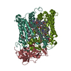


 PDBj
PDBj


 Assembly
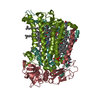


 Mass: 911.504 Da / Num. of mol.: 4 / Source method: obtained synthetically / Formula: C55H74MgN4O6
 Mass: 889.215 Da / Num. of mol.: 2 / Source method: obtained synthetically / Formula: C55H76N4O6
 Mass: 863.343 Da / Num. of mol.: 2 / Source method: obtained synthetically / Formula: C59H90O4
 Mass: 229.402 Da / Num. of mol.: 6 / Source method: obtained synthetically / Formula: C14H31NO / Comment: LDAO, detergent*YM
 Mass: 55.845 Da / Num. of mol.: 1 / Source method: obtained synthetically / Formula: Fe
 Mass: 94.971 Da / Num. of mol.: 1 / Source method: obtained synthetically / Formula: PO4
 Mass: 568.914 Da / Num. of mol.: 1 / Source method: obtained synthetically / Formula: C41H60O
 Sample preparation
 / Beamline: ID14-3 / Wavelength: 0.933 Å
 Processing